Download
1 / 33
330 likes | 475 Views
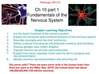
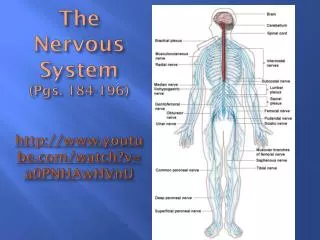
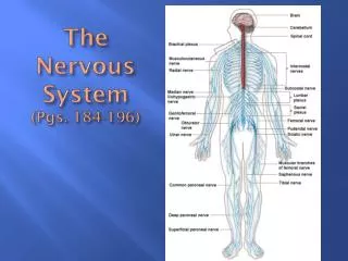
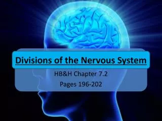
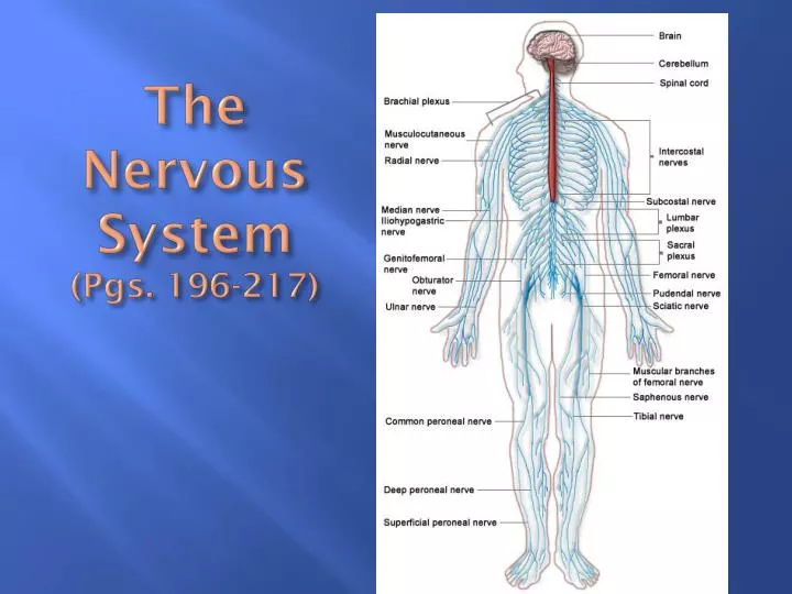
The Nervous System (Pgs. 196-217). The Nervous System. Organs of the nervous system are divided into Central Nervous System (CNS) Peripheral Nervous System (PNS). Vertebrate Nervous System. Central Nervous System Brain Spinal Cord

E N D
The Nervous System • Organs of the nervous system are divided into • Central Nervous System (CNS) • Peripheral Nervous System (PNS)
Vertebrate Nervous System • Central Nervous System • Brain • Spinal Cord • Peripheral Nervous system – cranial & spinal nerves & ganglia • Somatic (voluntary) – connect to skin and Skeletal Muscles • Autonomic (involuntary, homeostatic control) – cardiac muscle, smooth muscle, and glands • Sympathetic – dominates in times of stress; “fight or flight” syndrome (increases heart rate, blood pressure, breathing) • Parasympathetic – acts as counterbalance, conserves energy (decreases heart rate, blood pressure, breathing rate)
Central Nervous System • Brain – largest & most complex part of NS. Contains nerve centers associated with sensations. Issues motor commands & carries on higher mental functions: Fig. 8-9, 8-10
Brain stem • Extends from base of the cerebrum to the spinal cord • Consists of midbrain, pons, & medulla oblongata • Medulla oblongata – transmits all ascending & descending impulses, & contains several vital & nonvitalrelex centers (cardiac, respiratory, vasomotor) • Pons- transmits impulses between the cerebrum & other parts of the NS, and contains centers that help regulate the rate & depth of breathing • Midbrain – contains reflex centers associated with eye & head movements (visual & auditory impulses)
Diencephalon • Thalamus – central relay station for incoming sensory impulses; emotions and alerting or arousal mechanisms • Hypothalamus – maintain homeostasis, regulation of body temp., water balance, sleep-cycle control, appetite & sexual arousal (**very important gland of the endocrine system)
Cerebellum • Consists of two hemispheres that are connected by the vermis • Composed of white matter surrounded by a thing cortex of gray matter • Functions primarily as a reflex center in the co-ordination of skeletal muscle movements & the maintenance of equilibrium
Cerebrum • Two hemisphere connected by the corpus callosum • Surface marked by convolutions (ridges) and gyri (grooves); 4 lobes each hemisphere – frontal, parietal, temporal, occipital • Composed of thin layer of gray matter near surface; white matter found deeper • Higher brain functions such as thought, reasoning, interpretations of sensory impulses, control of voluntary muscles, and storage of memory
Spinal Cord • Nerve column that extends from the brain into the vertebral canal. It terminates at the level between L1 and L2; fig. 8-11, 8-12
Spinal Cord - Structure • Composed of 31 segments, each of which gives rise to a pair of spinal nerves • Characterized by two deep longitudinal grooves that divide it into right and left halves • Has a central core of gray matter that is surrounded by white matter • White matter is composed of bundles of myelinated nerve fibers
Spinal Cord - Function • Provides a two way communications system between the brain & body parts outside the NS • Primary reflex center • Ascending tracts carry sensory impulses to the brain; descending tracts carry motor impulses to muscles and glands • Many of the fibers in the ascending and descending tracts cross over in the spinal cord or brain
Coverings & Fluid spaces of the Brain & Spinal Cord • Meninges – protective membrane covering brain & spinal cord; three layers; Fig. 8-13 • Dura mater – outer layer • Arachnoid – middle layer • Pia mater – inner layer • Cerebrospinal fluid occupies the space between the arachnoid & pia mater
Coverings & Fluid spaces of the Brain & Spinal Cord • Ventricles – interconnected cavities within the cerebral hemisphere & brain stem that are filled with cerebrospinal fluid: Fig. 8-14
Peripheral Nervous System • Consists of cranial and spinal nerves that branch out from the brain and spinal cord to all body parts. Subdivided into somatic & autonomic portions.
Cranial Nerves Fig. 8-16, Table 8-2 • 12 pairs that connect the brain to parts in the head, neck, & trunk • Most cranial nerves are mixed (sensory & motor); some are pure sensory & other primarily motor • Some cranial nerve fibers are somatic & others are autonomic
Spinal NervesFig. 8-17, 8-18 • 31 pairs originate from the spinal cord • These mixed nerves provide a two way communication system between the spinal cord & parts in the arm, legs, neck, and trunk • Each nerve emerges by a dorsal and ventral root • Dorsal (posterior) root contains sensory fibers and is characterized by the presence of a dorsal root ganglion • Ventral root contains motor fibers
Spinal NervesFig. 8-17, 8-18 • Each spinal nerve divides into several branches & then combines with other spinal nerves to form plexuses in which nerve fibers are sorted and recombined so that those fibers associated with a particular part reach it together.
Somatic Nervous System • Skeletal muscle & skin • Conduction of nerve impulse is all the way from the spinal cord or brain to the effector • No synapses • Fig. 8-19 (left side of picture)
Autonomic Nervous System • Portion of nervous system that function without conscious effort • Concerned primarily with the regulation of visceral activities that aid in maintaining homeostasis • Fig. 8-19 (right side of picture) • Made up of motor neurons that conduct impulses to cardiac and smooth muscle tissue & glandular epithelial tissue
Autonomic Nervous System • Conduction pathway is a 2 neuron relay – in the ganglia the impulses are integrated before passing out to effectors • ANS is divided into 2 divisions • Sympathetic • Parasympathetic
Sympathetic • Prepares the body for stressful and emergency conditions “fight or flight syndrome” • Increase in heart rate, breathing, decrease in activities of digestive tract • Sympathetic preganglionic dendrites and cell bodies are located in the gray matter of the thoracic and upper lumbar segments of the spinal cord • Sympathetic preganglionic axons synapse with many postganglionic neurons in sympathetic ganglion & these postganglionic neurons frequently terminate in widely separated organs; therefore, sympathetic responses are usually widespread
Parasympathetic • Most active under ordinary conditions • Decreases heart rate and breathing • Opposite (antagonist) to sympathetic • Parasympathetic preganglionic dendrites and cell bodies are located in gray matter of brain stem and sacral segments of the spinal cord; axons extend some distance before terminating in parasympathetic ganglion • Parasympathetic postganglionic neurons have short axons that extend into nearby structures; therefore parasympathetic stimulation usually involved responses by only one organ
Neurotransmittters – chemical compounds released by axons • The different effects of the autonomic divisions are due to the different neurotransmitters released by the postganglionic fibers • There are various types of receptors present on the cell membranes and a cell’s response to neurotransmitters depends upon the number and type of receptors present in their membranes. • Fig. 8-20