Download
1 / 25
250 likes | 449 Views
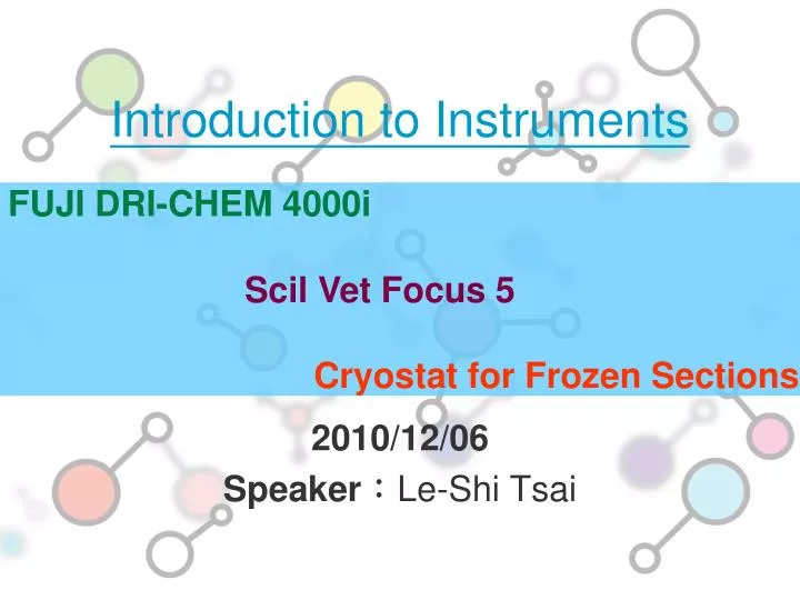
Introduction to Instruments. FUJI DRI-CHEM 4000i Scil Vet Focus 5 Cryostat for F rozen S ections. 2010/12/06 Speaker : Le-Shi Tsai. Notice for the use of animal center Instrument.

E N D
Introduction to Instruments FUJI DRI-CHEM 4000i Scil Vet Focus 5 Cryostat for Frozen Sections 2010/12/06 Speaker:Le-Shi Tsai
Notice for the use of animal center Instrument • Please attend illustration meeting before you begin operating instruments. If you do not participate in the illustration meeting , you may NOT use the instrument.(The meeting will be held 2-3 times per semester, and please pay attention to the animal center announcement.) • Please fill the registration forms 1 or 2 days prior to use the instrument to facilitate preparation.
FUJI DRI-CHEM 4000i Biochemistry Analyzer
The registration • forms • Exception code • table Need to purchase reagents by user Main instrument
Colorimetric Potentiometric Linear Measurement time
External Structure of the Instrument Light source 廢料盒 Can not open the lid whenindication light is on
Automatic detection 1. can be automatically diluted 2. A test only needs at least 10μL. ( Manual detection )
Principle:Colorimetric • Spreading layer: • filtered out macromolecules (proteins, dyes, etc.) • evenly spread the fluid sample • into the reaction layer β-D-glucose+O2+H2O GOD D-gluconic acid+H2O2 1,7-Dihydroxynaphthalene+4-aminoantipyrine+H2O2 POD Red dye 505nm reflection density calibration curve glucose concentration
Principle:Potentiometric generated voltage difference between ion selective electrodes applied to a reference solution and to the sample potentiometric difference between the sample and the reference solution is measured E = E0 + 2.303 * RT/Nf * Log ai ai = f * Ci Ci = Ion concentration
Scil Vet Focus 5 Hematology Analyzer
Introduction to Instrument • The following animals can be detected: dog、 rat、 pig、rabbit、mouse、 G. pig • The following items can be detected:WBC、DC、RBC、HGB、HCT、MCV、MCH、MCHC、RDW、Retics、PLT、MPV、PCT Slide 002
Equipment the registration forms the kit printer waste pail main Instrument
Equipment the kit 1.Diluent 2.Clean Deproteinizer 3.Cellyse 1.red blood cell lysis 2.reagent to convert all hemoglobin to the stable cyanmethemoglobin form
Principles • A distinguishing feature of blood cell types is • the diameter of the cells.(RBC、WBC、Platelet) • 2. WBCs are classified according to the characteristics of their cytoplasm and nucleus.3.Cyanmethemoglobin can then be detected by a spectrophotometer set at 540 nm. The concentration of hemoglobin is then calculated from the optical density of the solution. 感 應 區 The cells are forced to pass through aperture in single file (one by one). Cells are injected into the core of a sheath flow and confined to a narrow single-file stream by hydrodynamic focusing(15μm). Diluent form a vortex
Automatic Mode • open the lid • mix the specimen well • put your sample into the specimen-container housing rack
Leica CM3050 S Cryostat for Frozen Sections
Frozen Tissue Processing tissue process and cutting fixing and embedding only use O.C.T as embedding medium freeze and slice Hard tissue using a dedicated film stain mounting observe it under a microscope
The advantage of Leica CM3050 S • It is ideal for undecalcified hard tissue : 1.can be set to any section thickness and section automatically 2. with a dedicated film, can avoid the hard tissue broken. • with rough-cut function • adjustable specimen temperature: In accordance with the type of specimen to select the appropriate temperature. • adjustable cutting speed