Download
1 / 27
270 likes | 419 Views
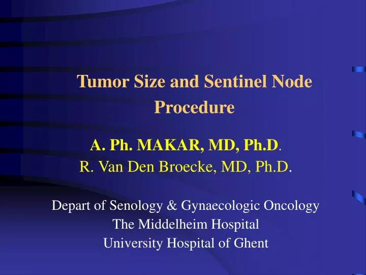
Tumor Size and Sentinel Node Procedure. A. Ph. MAKAR, MD, Ph.D . R. Van Den Broecke, MD, Ph.D . Depart of Senology & Gynaecologic Oncology The Middelheim Hospital University Hospital of Ghent. Tumor size. I . Carcinoma in situ II. T1 & T2 (<3cm ) tumors III. Large T2 & T3 tumors

E N D
Tumor Size and Sentinel Node Procedure A. Ph. MAKAR, MD, Ph.D. R. Van Den Broecke, MD, Ph.D. Depart of Senology & Gynaecologic Oncology The Middelheim Hospital University Hospital of Ghent
Tumor size I. Carcinoma in situ II. T1 & T2 (<3cm ) tumors III. Large T2 & T3 tumors IV. Inflammatory breast cancer • Multi-centric / multi-focal disease Prospective analysis Middelheim hospital 1998-2001, 268 patients, single surgeon
Sense of SN procedure • Impacton further surgical management, postoperative treatment or prognosis • False negative rate: acceptable • Number to be saved complete ALND: high • Number that needs second surgery: low • increased morbidity: swelling, numbness, pain • increased coasts • completeness of axillary dissection ?
I. Ductal Carcinoma In Situ • Silverstein: rate of axillary metastases < 1% • Survival rate > 98% • Axillary staging is generally not necessary • IHC: micro-metastases in 5-15% of cases • Lara (2003) & Broekhuizen & Marby (2006): • No impact on local failure or distant metastasis
ADH/DCIS in core biopsy: underestimation risk • Underestimation risk of invasive disease : 20-40% • SN procedure can be justified: • Mammographic lesion >5cm • Underlying mass/distortion • Palpable lesion & Core biopsy under sonography • High grade lesion & micro-invasion &LVSI
II. T1 &T2 tumors (<3cm) • Extensive evaluation: ASCO guide lines • Identification rate >95% • Failed identification: • Age >60 years • Capsular invasion, high number of positive nodes • FNR <10%: removal of all radioactive nodes • IHC: more micro-metastases 15% (10-67%) • SN metastases in <50% of tumors • SN only site of metastases in 40%
Positive SN macro-metas micro-metas Complete ALNDAlternatives ? ? RadiotherapyObservation (EORTC) ACSOG00Z11 Historical NSBAP-04
Micro-metastases in SN: Risk factors predicting Non-SN metastases • LVSI • Tumor size • Extra-nodal spread • Micro-metastasis: • Size of micro-metastasis • Micro-metastasis detected by HES vs IHC • Location of micro-metas: sinusal vs intranodal • Number of pos SN/total nr of SN: (1/3)
Rate of Non-SN involved in case of micro-metastases in SN according to tumor size
T1 tumors & micro-metastasis in SN Houvenaeghel (2006) & Leikola (2006): • pT1a, pT1b (IHC) • pT1a- pT1c of tubular, colloid or medullary types • Risk of Non-SN involvement: <5% • Risk of involvement of >1 Non-SN : 0% • ALND can be omitted with minimal risk
Prediction of Non-SN metastases in case of micro-metstases in SN • Turner (2000): likelihood model • Van Zee (2003): nanogram (9 variables) • Meta-analysis: • No combination of factors was able to predict non-SN metastases • 10% of the micro-metastases in the SN were associated with one or more macro-metastases in Non-SN
ALND dissection is recommended in every case with micro-metastases in the SN The prognostic significance micro-metastases: The Ludwig Breast Cancer Study Group NSABP-32 ACSOG Z0010
% patients with tumors > 3cm and pos SN that have an additional disease in Non-SN
SN with T3 tumors • The high risk of nodal metastases warrants complete ALND unless: • Motivated patient to have LN conservation
SN procedure following pre-operative CT: Meta analysis • Identification rate (IR): 91% • IR isotope 95% vs 93% blue dye • No serious concern regarding the fibrotic effect of CT on lymphatic pathways • False negative rate: 12%
Neo-adjuvant chemotherapy & axillary downstaging • Anthracyclin / cyclophosph based CT provides up to 30 % axillary down staging • Size of residual LN metastases after neo-adjuvant CT is of prognostic significance • Changing concept: • SN prior to neo-adjuvant CT followed by “2nd look” axillary dissection post CT = better prognostic information
Tumors >3cm with macro-metastases in SN = almost 100% non-SN metastases SN prior to CT (better staging) Axillary dissection post CT Pathologic remission Persistent disease Less morbidity
IV.Inflammatory breast cancer • Insufficient data. • High risk of nodal spread • False negative rate: • Occlusion of subdermal lymphatics (tumor emboli)
V. Multicentric tumors • Occurs in up to 10% of cases • Were excluded by most SN investigators • Hypothesis “sentinel for the entire breast”: • High success ratio • No increase in false negative ratio • Peri-areolar injection
Conclusions-1 • DCIS: • In some cases of core biopsy with risk of underestimation: • Lesions > 5cm • Underlying lesion: density/distortion • High grade tumors & micro-invasion, LVSI • Immediate reconstruction
Conclusions-2 • T1 –T2 (< 3cm): • Standard procedure with N0 • With few exceptions “T1a and T1a-T1c of certain pathology”, a full ALND is indicated in case of microscopic disease in the SN • The prognostic significance of micro-metastases needs further evaluation
Conclusions-3 • Large T2 & T3 tumors: • IR and FNR are comparable with T1 tumors • Yet the high incidence of LN metastases makes the clinical relevance of SN procedure of limited value except in case of neo-adjuvant CT • Multi-centric /multi-focal disease: • More reports suggest safety of the procedure • Yet multifocal tumors have higher risk of nodal spread than unifocal ones of same diameter
Conclusion-4 • 2nd axillary surgery carries more morbidity: Prospective multi-centric trial comparing immediate versus “second-look” axillary surgery post chemotherapy in patients with positive SN: Welcome to participate
Nuclear medicine: K. Melis F. Van Acker Pathology: S. Declercq L. Van Leuevn C.Mattelaer Radiotherapy: D. Van denWeyngaert S. Vanderkam I. Jacobs Medical Oncology E. Joossens D. Becquart A..Vandebroek Sentinel Node Team