Download
1 / 18
180 likes | 378 Views
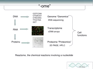
Harini Chandra Affiliations. Protein chemistry to proteomics. Technological advancements in protein analysis with increased sensitivity, resolution and capability to carry out high throughput studies has led to a transition from protein chemistry to the new field of proteomics.

E N D
Harini Chandra Affiliations Protein chemistry to proteomics Technological advancements in protein analysis with increased sensitivity, resolution and capability to carry out high throughput studies has led to a transition from protein chemistry to the new field of proteomics.
Master Layout (Part 1) 1 This animation consists of 3 parts: Part 1 – Mass spectrometric analysis Vs Edman degradation Part 2 – Immobilized pH gradient (IPG) strips Vs tube gels Part 3 – Completion of several genome sequence projects Sample inlet Charged peptide fragments 2 + Mass analyzer + + + + + + + + Intact protein to be analyzed Mass spectrometry 3 Detector Ionization source Edman degradation Labeling Labeling Release Polypeptide chain 4 Labelled amino acid Release 5
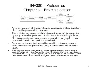
Definitions of the components:Part 2 – Mass spectrometric analysis Vs Edman degradation 1 1. Mass spectrometry:A powerful protein detection and analysis technique that produces charged molecular species in vacuum, separates them by means of electric and magnetic fields and measures the mass-to-charge ratios and relative abundances of the ions thus produced. 2. Protein to be analyzed: Mass spectroscopy is commonly used to identify proteins by breaking them into smaller, charged peptide fragments and analyzing their mass-to-charge ratio. Several advancements have been made in MS to facilitate this process. 3. Sample inlet: The first point of contact where the sample is introduced within the mass spectrometer either as liquid nano-droplets or alongside small matrix molecules. 4. Ionization source: The protein of interest must be ionized with a suitable source so that the charged molecules can be detected in the mass spectrometer. Biological samples are most often ionized by electrospray ionization (ESI), wherein liquid containing the analyte of interest is dispersed into a fine aerosol by means of an electrospray. The other commonly used technique is matrix assisted laser desorption/ionization (MALDI), another soft ionization technique that involves the use of a laser beam (nitrogen) for ionization. The biomolecule of interest is embedded in a solid matrix that prevents it from being damaged by the laser. 2 3 4 5
Definitions of the components:Part 2 – Mass spectrometric analysis Vs Edman degradation 1 5. Mass analyzer: The charged molecules are segregated after ionization based on their mass-to-charge ratio. The four most common analyzers are Time of Flight (TOF), quadrupole, ion trap and Fourier Transform Ion Cyclotron Resonance (FT-ICR), each having its own level of sensitivity and accuracy. The ion motion is manipulated either by electric or magnetic field and ions are directed to the detector for analysis. 6. Charged peptide fragments: The peptide fragments generated by the ionization source carry positive, negative as well as neutral charges. The settings of the mass analyzer can be adjusted such that only certain ions are taken up for detection. A cut off range can be specified whereby only ions in that particular range move ahead for detection. Sensitivity of detection for positive ions is higher than negative ions while neutral ions cannot be detected by MS. 7. Detector: The final component of the spectrometer is the detector which can record either the current produced or the charge induced when an ion hits a surface. Electron multipliers are commonly used as detectors. 8. Edman degradation: This is a technique for sequencing amino acid residues in a polypeptide chain starting from the N-terminus without disrupting any other peptide bonds. The peptide is treated with phenylisothiocyanate and then hydrolyzed such that only the derivatized amino acid is liberated, leaving the remaining peptide chain intact for the next round of analysis. Although this is an extremely useful technique, it is time consuming and cumbersome. 2 3 4 5
Part 1, step 1: 1 The development of these soft ionization techniques which could be used for protein samples was a major turning point for proteomic studies. Koichi Tanaka and John Bennett Fenn were awarded the Nobel Prize in Chemistry in 2002 for development of these techniques! 2 Sample ionization Laser beam 3 Electron beam ESI MALDI 4 Action Description of the action Audio Narration Small circles must be show to enter tube and then the coil must be zoomed into to show the figures below. First show some coloured circles entering the transparent tube on top and moving towards the grey coil. This is then zoomed into and the figures below must be shown with the circles moving towards the red rectangle at the end. The text box on right top must appear followed by the bubble on top left. Protein analysis by MS had proved challenging due to complete degradation of samples with then available hard ionization techniques. This was overcome by development of soft ionization techniques, MALDI and ESI. The vapourized sample is ionized by means of an electron beam in ESI or by a laser beam in MALDI. This results in charged peptide fragments which get accelerated towards the mass analyzer. These two techniques greatly impacted proteomic studies as they facilitated MS analysis of protein samples. 5
Part 1, step 2: 1 Edman degradation Release Release …... Polypeptide chain Labeling Labelled amino acid 2 Labelled amino acid Multiple rounds of sequencing required to analyze long polypeptide chains by Edman degradation. Sequencing by mass spectrometry is rapid thereby facilitating analysis of large number of samples. Mass spectrometry 3 + Sample inlet + + Charged peptide fragments + + + + + + Rapid sequencing Intact protein to be analyzed MS data analysis Mass analyzer Detector 4 Ionization source Action Description of the action Audio Narration (Please redraw all figures.) As shown in animation. First show the chain with circles on top. The 1st circle must be attached to a triangle as shown. This must then be released after which the second circle is attached to the triangle. Again this is released leaving behind the remaining circles after which the dots must appear to show that this process continues. Next the figure below must be shown. The purple ‘protein’ must enter via the inlet and arrive at the grey circles. Here, it must be broken down into smaller fragments as shown. The smallest green fragments must travel rapidly to the ‘detector’ followed by the red fragments and finally the violet fragments which must move slowly. Next, the arrow and the computer must appear followed by the text box shown. Protein sequencing by Edman degradation is time-consuming and cumbersome. Several rounds of sequencing are required for analysis of long polypeptide chains. Protein sequencing by MS, however, is much faster thereby allowing large number of samples to be analyzed in same amount of time. 5
Master Layout (Part 2) 1 This animation consists of 3 parts: Part 1 – Mass spectrometric analysis Vs Edman degradation Part 2 – Immobilized pH gradient (IPG) strips Vs tube gels Part 3 – Completion of several genome sequence projects 2-D Gel electrophoresis Separation based on pI 2 IPG strip Tube gel 3 Separation based on molecular weight 4 SDS-polyacrylamide gel 5 Source: Biochemistry by A.L.Lehninger, 4th edition (ebook); http://www.gelifesciences.com/
Definitions of the components:Part 2 – IPG strips Vs tube gels 1 1. 2-D gel electrophoresis: This is an advanced electrophoretic separation technique that carries out separation in two dimensions. Proteins are separated based on their isoelectric points (pI) in the first dimension followed by SDS-PAGE in the second dimension, which separates proteins based on their molecular weight. 2. IPG strip: Commercially available immobilized pH gradient (IPG) gel strips have considerably facilitated the process of isoelectric focusing by eliminating the tedious steps of gel preparation and pH gradient establishment using ampholyte solutions. These strips, available across the pH range, contain a preformed pH gradient immobilized on a precast polyacrylamide gel placed on a plastic support. Narrow pH ranges can be selected for fine separations while broader pH ranges are also available for crude separations. These strips only need to be rehydrated with a suitable buffer before use. 3. Tube gel: Isoelectric focusing using tube gels is a tedious process compared to the readily available IPG strips. Here, the gels first need to be cast and then run with a suitable ampholyte solution before sample application, in order to establish the pH gradient. These pH gradients are not very stable and tend to breakdown on application of some concentrated samples. 4. SDS-polyacrylamide gel: It is one of the most commonly used gels for separation of proteins based on their molecular weights. It is prepared by free radical induced polymerization of acryl-amide and N, N’-methylenebisacrylamide and also contains the anionic detergent, sodium dodecyl sulhpate (SDS), and reducing agent, dithiothreitol (DTT). These are responsible for denaturing the protein and facilitate their separation solely on the basis of size. 2 3 4 5
Part 2, step 1: 1 2-DE of same protein sample carried out using tube gels Increasing pH pH 9 pH 3 Tube gel 2 Decreasing molecular weight 3 SDS-PAGE 4 Action Description of the action Audio Narration As shown in animation. (Please redraw all figures.) First show the tube on top with the bands followed by the empty blue slab below. The tube must then move down and get fixed n the groove of the slab. The blue spots must then appear which move down the blue slab in the direction indicated until they reach the positions shown. This is repeated for the remaining two slabs as well. The pH gradient in tube gels is established by means of ampholyte solutions which consist of low molecular weight organic acids and bases that are subjected to an electric field. These gradients are not always very stable and tend to break down upon addition of concentrated samples. Analysis of the same protein mixture by 2-DE using tube gels often gives a lot of variation in results across gels. 5
Part 2, step 2: 1 Reproducibility across gels is not obtained in 2-DE using tube gels. Commercially manufactured IPG strips ensure minimal variations in the pH gradient of the strip thereby giving better reproducible results. 2-DE of same protein sample carried out using IPG strips Increasing pH Increasing pH Increasing pH pH 9 pH 9 pH 9 pH 3 pH 3 pH 3 2 IPG strip Decreasing molecular weight 3 SDS-PAGE 4 Action Description of the action Audio Narration As shown in animation. (Please redraw all figures.) First show the strip on top with the bands followed by the empty blue slab below. The tube must then move down and get fixed n the groove of the slab. The blue spots must then appear which move down the blue slab in the direction indicated until they reach the positions shown. This is repeated for the remaining two slabs as well. The problem of reproducibility has been overcome to a large extent by the development of IPG strips which are commercially manufacture gel strips having a preformed pH gradient. These strips only need to be rehydrated before use for 2-DE. Minimal gel-to-gel variation is observed when the same sample is run by 2-DE using IPG strips, thereby making them extremely suitable for large scale proteomic applications. 5
Master Layout (Part 3) 1 This animation consists of 3 parts: Part 1 – Immobilized pH gradient (IPG) strips Vs tube gels Part 2 – Mass spectrometric analysis Vs Edman degradation Part 3 – Completion of several genome sequence projects Genomic DNA Digestion & insertion into BACs 2 Contigs identified & mapped 3 Sequencing 4 Sequence overlaps reveal final sequence 5 Source: Biochemistry by A.L.Lehninger, 4th edition (ebook)
Definitions of the components:Part 3 – Completion of several genome sequence projects 1 1. Genomic DNA: The entire DNA sequence of all chromosomes of an organism constitutes its genomic DNA. There have been several projects aimed at deciphering the complete genome sequence of organisms including humans. 2. BAC: Bacterial Artificial Chromosomes are DNA constructs that are useful for cloning purposes. These cloning vectors can carry DNA inserts of around 150-350 kbp and have been extremely useful in the various genome sequencing projects carried out. 3. Contigs: A set of overlapping DNA fragments that are obtained from a single genetic source. These contigs are used to deduce the original DNA sequence. 4.Sequencing: DNA fragments that have been amplified using the BAC are sequenced to obtain the base pairs of each fragment. These are then used to deduce the original sequence of the intact DNA by aligning the fragments having overlapping end sequences. 2 3 4 5
Part 3, step 1: 1 Fragmentation & insertion into BAC 2 Genomic DNA Restriction endonuclease Contigs aligned 3 Sequence alignment Final DNA sequence 4 Action Description of the action Audio Narration The pie-shaped object must break the blue line into several small pieces. (Please redraw all figures.) First show the blue line on the left top. The pie-shaped object must then move along this blue line and cut it up into smaller pieces. These must then be straightened out and aligned next to each other. The sequence must then be shown to appear with the pink regions highlighted followed by the final sequence shown above. The genomic DNA is cleaved using a suitable restriction endonuclease and inserted into the bacterial artificial chromosome. The amplified sequences are sequenced using an automated sequencer and then mapped by aligning the overlapping fragments to obtain the original DNA sequence. 5 Source: Biochemistry by A.L.Lehninger, 4th edition (ebook)
Part 3, step 2: 1 GCCTTTAAGGATCCGGA TTGCAAATCCCGATTCG DATABASE SEARCH – Corresponding protein sequence obtained from genomic sequences GGCGTTATGGCTTGGAA 2 CCCGGATGCCTGGTCCA 3 Available genome database MS sequencing Ala-Leu-Val-Cys-Trp-Tyr-Ala-Gly-Gly-Tyr-His-Pro-Met-Arg-Ile-Lys-Lys-Glu-Ser-Pro-Thr-Thr-Val-Val-Gln Protein Protein sequence 4 Action Description of the action Audio Narration First show the computer screen with all the sequences. Next show the green protein below followed by the ‘MS’ apparatus. The protein must be shown to move through the tube and from the other end, the sequence shown must appear. This sequence must then be shown to enter the computer screen and the green star with text must appear out of it. Genome sequences of several organisms, including humans, have been successfully completed and these genome databases are extremely useful in correlating gene and protein sequences. Several databases are now readily available which can easily help in identifying gene sequence of a protein that has been sequenced by mass spectrometry. As shown in animation. 5
Interactivity option 1:Step No:1 1 A genomic DNA sequence was cut into 5 fragments using a suitable restriction endonuclease. After amplification in the BAC, sequencing of these fragments gave the following sequences: 1. ATTTGCAAATCCCGGAAT 2. GCCTTTAAGGATTTG 3. GGAATCCCGGATGCCTATAT 4. CGGGCGTTATGGCTTAC 5. TATATGGCCCAATACGCGGGC What is the sequence of the intact original DNA? 2 A) GCCTTTAAGGATTTGCAAATCCCGGAATCCCGGATGCCTATATGGCCCAATACGCGGG CGTTATGGCTTAC B) ATTTGCAAATCCCGGAATGCCTTTAAGGATTTGGGAATCCCGGATGCCTATATCGGGCGTTAT GGCTTACTATATGGCCCAATACGCGGGC 3 C) GCCTTTAAGGATTTGCAAATCCCGGAATTATATGGCCCAATACGCGGGCGTTATGGCTTACGG AATCCCGGATGCCTATAT D) GCCTTTAAGGATTTGCAAATCCCGGAATGGCCCAATACGCGGGCGTTATGGCTTACCCC GGATGCCTATAT 4 Results Interacativity Type Options Boundary/limits User should be allowed to choose one of the four options but can continue to choose until he arrives at the right answer. The correct answer (A) must turn green if chosen while remaining must turn red if selected. User should be directed to next slide, step 2, upon choosing correct answer. Choose the correct option. 5
Interactivity option 1:Step No:2 1 By analyzing the end sequence overlaps and aligning the fragments, the entire original DNA sequence can be deduced. GCCTTTAAGGATTTG ATTTGCAAATCCCGGAAT 2 GGAATCCCGGATGCCTATAT TATATGGCCCAATACGCGGGC CGGGCGTTA-TGGCTTAC 3 GCCTTTAAGGATTTGCAAATCCCGGAATCCCGGATGCCTATATGGCCCAATACGCGGG CGTTATGGCTTAC 4 5
Questionnaire 1 1. Which of the following reagents is used for Edman degradation process? Answers: a) Dansyl chloride b) Urea c) Phenylisothiocyanate d)Methyisothiocyanate 2. Which of the following is not a mass analyzer in MS? Answers: a) TOF b) MALDIc) Quadrupole d) Ion trap 3.What pH range can be used to separate a crude protein extract containing 100 proteins of varying isoelectric points? Answers: a) pH 2-3 b) pH 4-5 c) pH 8-9 d) pH 3-11 4.There are three fragments A, B and C of lengths 30, 40 and 50 nucleotides respectively. The region of overlap between fragment A and B is of 15 nucleotides while that between A and C is of 10 nucleotides. What is the total length of the intact DNA segment? Answers: a) 100 b) 95 c) 120 d) 85 2 3 4 5
Links for further reading Books: Discovering Genomics, Proteomics & Bioinformatics, 2nd edition, A.Malcolm Campbell & Laurie J.Heyer Research papers: Gorg, A. et al. The current state of two-dimensional electrophoresis with immobilized pH gradients. Electrophoresis 2000, 21, 1037-1053. Gorg, A. et al. Two-dimensional polyacrylamide gel electrophoresis with immobilized pH gradients in the first dimension (IPG-Dalt): the current state of the art and the controversy of vertical versus horizontal systems. Electrophoresis 1995, 16, 1079-1086. Gorg, A et al. 2-DE with IPGs. Electrophoresis 2009, 30, S122-S132.