Download
1 / 46
460 likes | 578 Views
C4-11M B Cell Activation and Function III Immunity Mediated by B Cells and Antibody. Reading: Parham: pages 159-182 Michael Wolcott December 3, 2001. School’s Out The B Cell Goes to Work. B-Cell Development. Bone Marrow. Bone Marrow. Periphery. Periphery. Y. Y. Y. Y. Y. Antigen.

E N D
C4-11M B Cell Activation and Function IIIImmunity Mediated by B Cells and Antibody Reading: Parham: pages 159-182 Michael Wolcott December 3, 2001
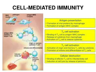
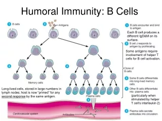
B-Cell Development Bone Marrow Bone Marrow Periphery Periphery Y Y Y Y Y Antigen IgM IgD Y Y Y Y IgM Y Y Y Y Plasma Cell Immature B Cell Omnipotent Stem Cell Mature B Cell Pro- B Cell Lymphoid Stem Cell Pre- B Cell Y Activated B Cell Memory B Cell Antigen Independent Antigen Dependent Primary Lymphoid Organ Secondary Lymphoid Organs
Antigen Driven B Cell Development Y Y Y Y Periphery Periphery Y Y IgM Y Y Y Y Y Y Antigen Y Y IgM Y Y Y Y Plasmablast Plasma Cell Class Switch Hypermutation Mature B Cell Activated B Cell Y Y Memory B Cell Antigen Dependent Secondary Lymphoid Organs
CORE MATERIAL • Induction of activation and proliferation of B cells, by most antigens, especially proteins, requires both binding of the antigen by the BCR and interaction of the B cell with antigen-specific activated helper T cell. • Armed helper T cells activate B cells that recognize the same antigen. • B-cell responses to bacterial polysaccharides do not require peptide-specific T-cell help. • Binding of antigen by the BCR leads to clustering of the receptors. Clustering of the antigen receptors leads to activation of intracellular signal molecules and internalization and processing of the antigen. • Germinal center B cells undergo V region somatic hypermutation and cells with mutations that improve affinity for antigen are selected. • Isotype switching and affinity maturation of B cells requires T cell help. • Formation of memory B cells requires T cell help. • Resolution of an infection is accompanied by death of most of the effector cells and the generation of memory cells.
Two Signal Model for B Cell Activation Competence Signal Progression Signal
The Initial Stages of Signal Transduction by an Activated B Cell ReceptorSignal 1
Co-Receptors on B Cells • The yang – CD19-CD21-CD81 complex co-stimulates signaling through the BCR. – couples the innate immune recognition of microbial antigens by the complement system to the activation of B-cells. • The yin- CD22 and FcRIIb1 have ITIMs (immunoreceptor tyrosine based inhibitory motifs) and down regulate the signaling through the BCR.
Co-receptorsSignal 2 • The yang – CD40-CD40 ligand interaction; cytokine receptors, B7/CD28, etc. • The yin- FAS-FAS ligand interaction inducing apoptosis.
Co-receptors-Signal 2 – The Yin • Lymphoid and other cells express Fas, and in lymphoid cells the levels of this protein increase upon antigen stimulation . • FAS ligand is a protein that is expressed mainly on activated T cells. • Fas ligand induced on helper T cells can recognize Fas on B cells and function to limit antibody responses, especially if the B cells are not specifically protected by antigen recognition and the induction of anti-apoptotic factors. • The interaction between Fas and Fas ligand induce apoptosis in the Fas expressing B cell that is not expressing anti-apoptotic factors.
Fundamental ConceptsImportance of Ag Structure on BCR Signaling
Shaping of the Repertoire • Somatic Hypermutation • Affinity Maturation = mutation and selection. • Class Switching
Somatic HypermutationFine Tuner of Humoral Immune Responses • Occurs in the germinal centers • Switched on after initial antigen-induced proliferation • Rate of mutation very high • Mutations are not radomly distributed • Localized to V region and flanking sequences • Include hot spots • Resulting clones which have higher affintiy for antigen tend to be selected into the memory pool
Isotype SwitchingRecombination Between Specific Switch Regions
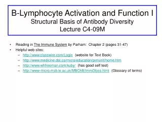
The Humoral Immune Response • The kinetics of antibody formation and other properties of the primary and secondary immune responses differ. • Primary – long lag period; IgM first antibody to appear followed by a gradual switch to other classes; low affinity ab. • Secondary – short lag period; more rapid rise in antibody titer; IgG and other classes predominate over IgM; higher affinity ab.
Comparison of Primary and Secondary Immune Response 2nd Immunization 1st Immunization Primary Response Secondary Response IgG Antibody Concentration Lag IgM Lag Time After Immunization
The Specificity of Immunologic Memory Antigen A + Antigen B Antigen A Secondary anti- A response Primary anti-A response Antibody Concentration Primary anti-B response Time After Immunization
Maturation and Clonal Selection of B Lymphocytes Mature B cells A B C d E C A x D X Clonal Selection Antigen X B cell activation Clonal Expansion B cell proliferation Cell Death B cell Differentiation Plasma Cells Memory Cell Memory Cell Y Y Anti-X antibodies Y Y Y Y Y Y Y
CONCEPT: In the defense against disease, antibodies generally do not kill or remove pathogens solely by binding to them. • Antibodies not only must recognize antigen, but also must invoke responses – effector functions – that will remove the antigen and kill the pathogen. • Variable regions of antibody are the sole agents of binding to antigen. • The heavy chain constant region (CH) is responsible for a variety of collaborative interactions with other proteins (e.g. complement), cells (elements of innate immune system), and tissues that result in the effector functions of the humoral response. • Not all classes of immunoglobulin have the same functional properties.
EFFECTOR FUNCTIONS OF ANTIBODIES • OPSONIZATION – the promotion of phagocytosis of antigens by macrophages and neutrophils. Protein molecules called Fc receptors (FcR), which bind the constant region of most classes of antibody are present on the surfaces of phagocytes. • ACTIVATION OF COMPLEMENT – IgM (most effective) and most subclasses of IgG can activate complement (Lecture C4-11). • ANTIBODY-DEPENDENT CELL-MEDIATED CYTOTOXICITY (ADCC) – The linking of antibody bound to target cells (virus infected cells, or some tumor cells) with FcR of natural killer cells (NK cells), neutrophils, macrophages,or eosinophils can result in killing of the target cell. • NEUTRALIZATION OF TOXINS AND VENOMS • NEUTRALIZATIONOF VIRUSES AND BACTERIA PREVENT ATTACHMENT TO CELL RECEPTORS
Neutralization by IgG Antibodies Protects Cells from Toxin Action
Viral and Bacterial Infection of Cells can be Blocked by Neutralizing Antibodies
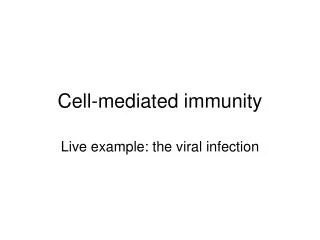
EFFECTOR FUNCTIONS OF ANTIBODIES • TRANSCYTOSIS • The delivery of antibody to the mucosal surfaces of the respiratory, gastrointestinal, and urogenital tracts, as well as its export to milk, requires the movement of immunoglobulin across epithelial layers and is mediated by the polyimmunoglobulin receptor. Although IgM can be transported to mucosal surfaces, IgA is the major antibody isotype found in secretions. • The transfer of IgG from mother to fetus requires that antibody must be transported across the placental tissue that separates the mother and fetus. This process is mediated by an FcRB (Brambell Fc receptor). • The transport of IgG from blood into extracellular spaces is mediated by the Brambell Receptor (FcRB) present on endothelial cells.
Transcytosis of Dimeric IgA Antibody Across Epithelia is Mediated by Poly-Ig Receptor
FcRB Transports IgG from Bloodstream into Extracellular Spaces Note: FcRB has a structure similar to MHC class I; i.e., is made up of an α-chain and β2 microglobulin. Note: FcRB is the same receptor that transport IgG across the placenta from mother to fetus.
In the First Year of Life Infants Have a Transient Decrease in Levels of IgG