Download
1 / 10
140 likes | 867 Views
Pulmonary Valve Stenosis. Bria Peacock. What is the Pulmonary Valve?. It is a valve that separates the right ventricle from the pulmonary artery. Through it, deoxygenated blood flows to the lungs by way the pulmonary artery to receive oxygen.

E N D
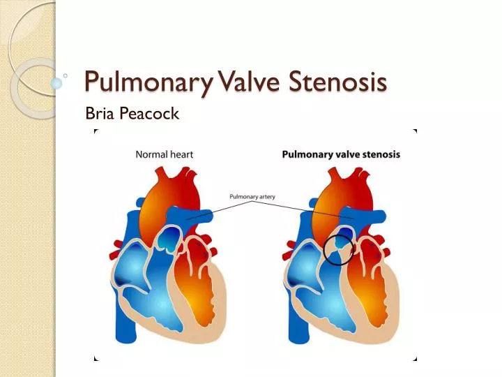
Pulmonary Valve Stenosis Bria Peacock
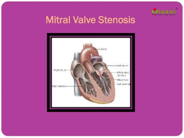
What is the Pulmonary Valve? • It is a valve that separates the right ventricle from the pulmonary artery. • Through it, deoxygenated blood flows to the lungs by way the pulmonary artery to receive oxygen. • The now oxygenated blood then flows back to the heart and then to the rest of the body.
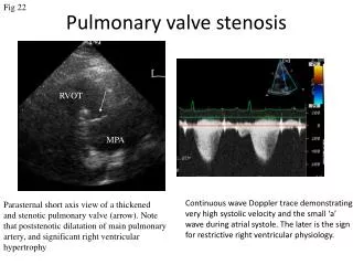
Pulmonary valve stenosis • The valve is narrowed in this disorder. • Because it does open wide enough, less blood gets to the lungs.
Symptoms • Abdominal swelling • Bluish color to the skin • Chest pain • Fainting • Fatigue • Poor weight gain • Shortness of breath • Sudden death
Causes • Birth defect • Cause unknown
Frequency • Rare • May run in families—genetic
Risk Factors • Family history of heart defect • Chromosomal disorders • Heart defects • History of pregnancy with fetal heart abnormalities or miscarriage • Viral infection during pregnancy
How is the disease identified • Heart murmur when listening to heart • Chest x ray • ECG—electrocardiogram which records the electrical activity of the heart • Echocardiogram creates a picture of the heart with sound waves • MRI or the heart creates a picture of the heart with magnets and radio waves • Cardiac catheterization is when a catheter is guided into either side of the heart from the groin or arm
Treatment • If mild, treatment is probably not needed • Medications could be used to treat the defects caused such as preventing clots with blood thinners and treating abnormal rhythms • Percutaneous balloon pulmonary dilation is done to widen the valve • Heart surgery
References • http://www.nlm.nih.gov/medlineplus/ency/article/001096.htm • http://www.med.nyu.edu/content?ChunkIID=618445#risk