Download
1 / 15
170 likes | 270 Views
Laser Microdissection. Introduction to LMD Technology Patrick Wojtkiewicz, Ph.D. North Louisiana Crime Lab (318) 227-2889 pwojtkie@NLCL.org. Microscope. Basic Microscopy. Terminology

E N D
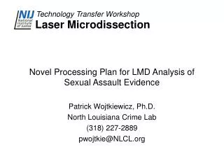
Laser Microdissection Introduction to LMD Technology Patrick Wojtkiewicz, Ph.D. North Louisiana Crime Lab (318) 227-2889 pwojtkie@NLCL.org
Basic Microscopy • Terminology • Magnification - apparent increase in size of the object, based upon microscope’s objective, ocular, and body tube. • Refraction – change in direction (velocity) of light as it passes from one medium to another (refractive index) • Illumination – method of applying light to the sample from the light source. • Köhler (Brightfield) • Vertical illuminator (Fluorescence)
Basic Microscopy • Brightfield • Light source is focused through sample & collected by objective lens. • Specimen refracts light, which bends it out of the collection angle aperture of the objective lens(dark). • Objective lenses most important part of microscope
Basic Microscopy • Fluorescence • Illuminate sample with UV (365 nm) light source (excitation) • Fluorochromes are energized and electron moves to higher energy state. • The energetic electron then drops back to its normal state releasing energy as a photon (emission). • Bypass filters block excitation , allowing emission (>420 nm)) to pass.
Basic Microscopy • Terminology • Resolution – the ability to distinguish two points as distinct and separate (~1000×NA). • Empty magnification is when magnification exceeds useful resolution
Basic Microscopy • Differential Interference Contrast • Optical method of creating relief (3-D) effects from a sample which has little inherent contrast. • Interference within the recombined beams causes effect. • Image formed by differences in the optical path caused by the specimen’s refractive index difference from the medium. • Note: Effect may be caused by a discontinuity in the film; therefore, interpretations should be made with care.
Pulse Lasers and Ablation • Ultrafast pulse lasers work by a multi-photon nonlinear process. • Photons of the ultrafast pulse are bunched at a very high density and material absorbs several photons at once. • Multi-photon absorption works in transparent media, such as plastics.
Pulse Lasers and ablation A nanosecond laser pulse first heats the target then melts it, then vaporizes it. The surroundings are heated as well leading HAZ problems. A picosecond laser pulse heats up material much faster. Heat has no time to spread. The material has no time to melt – it becomes plasma.
Advantages of Fast Pulse Lasers • The fast laser pulse (1.0 – 4.0 ns) is greater the heat diffusion time (~0.5 ns). • Material briefly melts – then energy strips electrons from molecules (plasma). • Nearly all of the energy of the pulse is used in plasma formation, not heating surrounding material. • Little debris or recast material. • Little collateral damage to surrounding area.
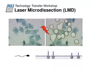
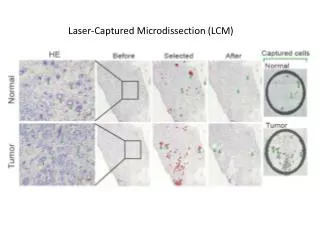
Laser Microdissection • History • Relatively brief history for forensics but well established in the medical field • First occurrence of laser microdissection was in 1996 • Primarily used to excise individual cells or groups of cells embedded in other tissue • First report on its forensic applications was at the AAFS meeting in 2004 but was initially used in 2003.
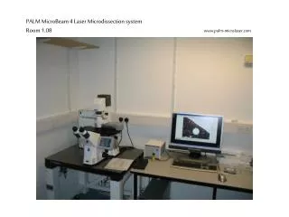
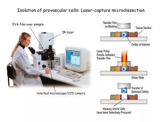
Types of LMD Microscopes • Leica • Automated microscope • Brightfield, fluorescence, alternative optics • Slides have film coating • Laser cuts plastic film, which allows the cut sample to drop into a tube. Up to four different samples can be placed on stage. • UV laser does cutting (355nm diode, 80 hz; 337nm gas, 30 hz). • Laser beam is directed though objective by prisms.
LCD Microscopes • Molecular Machines & Industries CellCut • Samples collected on a sticky cap • Samples can be easily inspected after cutting • Pulse laser cutting by moving stage • Contamination free