Download
1 / 70
710 likes | 852 Views
Nutrition, Environment, and Autoimmunity. Sabrina Schmitz EPOB 4800 Spring 2004. OUTLINE. REVIEW: IMMUNE SYSTEM “SELF” VS. “NON-SELF” DISTINCTION DEVELOPMENT OF AUTOIMMUNITY AUTOIMMUNE DISEASE MODELS ENVIRONMENT AND NUTRUTION CONCLUSION. Review. Lymphocyte Differentiation. Review.

E N D
Nutrition, Environment, and Autoimmunity Sabrina Schmitz EPOB 4800 Spring 2004
OUTLINE • REVIEW: IMMUNE SYSTEM • “SELF” VS. “NON-SELF” DISTINCTION • DEVELOPMENT OF AUTOIMMUNITY • AUTOIMMUNE DISEASE MODELS • ENVIRONMENT AND NUTRUTION • CONCLUSION
Review LymphocyteDifferentiation
Review • T cells: lymphocytes with antigen-specific receptors; play a major role in initiation and suppression of cell-mediated and humoral immunity • Cytotoxic T cells • Helper T cells
Review • B cells • Produce immunoglobulins (Ig): • B cell receptors: bound to B cell surface; allow antigen recognition (subsequent signaling or proliferation) • Secreted antibodies: unbound: “opsonize” pathogens. “Eat Me”
Cytokines and Chemokines • Cytokines: locally acting tissue hormones responsible for growth, differentiation, function, inhibition and apoptosis of cells. • Designations: type I, II; pro-inflammatory. • Ex. Interleukins, Interferons • Chemokines: chemo-attractant cytokines—very large and very diverse group of cytokines • Capable of inducing directional migration of activated leukocytes • mediate acute and chronic inflammation • Also regulate: cell-to-cell adhesion, angiogenesis, embryogenesis, etc. • Ex. Leukotrienes, platelet activating factor, transforming growth factor beta superfamily members, etc.
Apoptosis • Apoptosis is a genetically-regulated programmed cell death “program” in which the dying cell actively executes the decomposition and packaging of its internal contents in response to a variety of stimuli. It varies from necrosis in many ways, but most importantly, by its failure to induce an inflammatory response
Apoptotic pathways CD95
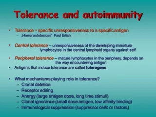
Development of autoimmunity *Dysregulation of apoptosis *Distinction between “self” and “non-self” blurred
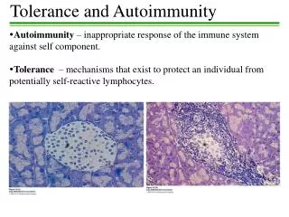
Distinction between “self” vs. “non-self” • Immune system must be able to distinguish between pathogenic cells and host cells • T and B cells have antigen-specific receptors • Autoreactive TCRs and BCRs must be deleted from repertoire. • Other lymphocytes take cues from T and B cells
T and B cell receptor diversification • The TCR and BCR repertoire of an individual must technically be able to identify every foreign antigen. • Several genetic recombination mechanisms are involved in diversification.
Filtration of autoreactive cells • Lymphocytes within the thymus are then given a “two signal test” • The receptor must: • Be able to recognize a foreign antigen in the context of an antigen presenting cell. • Not react with any autoantigens being presented to it (dendritic cells).
Antigen presentation HLA: human leukocyte antigen; various cell-surface proteins coded for by genes on the MHC; often necessary to activate major immune responses as a secondary safety mechanism
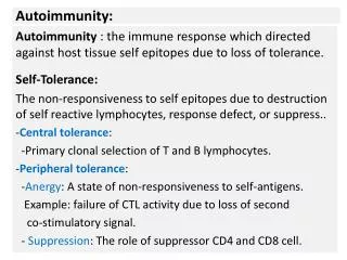
Definitions • Autoimmunity: state in which the immune system recognizes and removes host cells. • Autoimmune Disease: state in which autoreactive lymphocytes are allowed to proliferate and do harmful damage.
Development of autoimmunity • Three requirements • Genetic: • the individual must be able to produce HLAs capable of expressing autoantigens as well as TCRs and BCRs capable of recognizing them. • Additional genetic predispositions: faulty transcrption/translation enzymes, or tissue weakness. • Developmental • The tolerizing mechanisms must overlook the autoreactive lymphocytes (anergy, silencing) • Environment • An environmental trigger of some sort must initiate the reaction
Specific theories of autoimmunity development • Molecular mimicry: a structural similarity between a pathogen or environmental agent and a host antigen results in an immune response against both epitopes. • Cryptic epitope exposure: through alteration of a host antigen, it becomes structurally different enough to be recognized as a foreign invader
Once initiated, autoimmunity must propagate and “flare” to be considered an autoimmune disease
Once initiated, autoimmunity must propagate and “flare” to be considered an autoimmune disease
Once initiated, autoimmunity must propagate and “flare” to be considered an autoimmune disease
Autoimmune disease models Prevalence of various autoimmune diseases
Table***: Autoimmune diseases associated with targeted cell destruction (Kalden, 2003) DISEASE CELL KILLED Diabetes (IDDM) pancreatic β cell Multiple Sclerosis (MS) oligodendrocyte Hashimoto’s thyroiditis thyrocyte Sjögren’s syndrome acinus and ductal cells Polymyositis myocyte Primary biliary cirrhosis bile duct cells Autoimmune diseases associated with targeted cell destruction • Tissue-specific autoimmune diseases • Also, symptom-specific.
Ex. Multiple Sclerosis • a chronic demyelinating disease with observed onset usually in early adulthood and ultimately resulting in patches of hardened tissue in tissues • Damage to the central nervous system in MS is T cell mediated, and usually waxes and wanes throughout the lives of afflicted individuals. • Although no single unifying cause can be identified for MS development in all patients, the role of viral molecular mimicry is becoming increasingly emphasized in research. • Because both MS patients and healthy individuals express myelin basic protein (MBP) in the thymus, T cells must be rendered tolerant to the dominant epitopes of the protein. This is strong evidence for molecular mimicry and exposure of cryptic epitopes in the development of MS (Paul, 1999). • The most popular theory: “only during episodes of peripheral T-cell activation by crossreactive viral epitopes do these normally quiescent lymphocytes acquire the capacity to traverse the blood-brain barrier and initiate the immune response against myelin in the central nervous system” (Paul, 1999).
Other disorders involving CNS autoimmunity • MS, Alzheimer’s Disease (AD), Parkinson’s Disease (PD), amyotrophic lateral sclerosis (ALS), and to a certain extent, mental disorders (schizophrenia, bipolar disorder (BPD), attention deficit and hyperactivity disorder (ADHD), and depression) can be categorized as diseases associated with target cell death. • Recent research has begun looking at the etiologies neuropsychiatric disorders from a common mechanistic perspective: CNS autoimmunity and/or neuronal apoptosis. Specific points of interest: • Nerve growth factor (NGF) is a trophic factor, responsible for localized inhibition and proliferation of neurons during the development of the nervous system. In the absence of this neurotrophin, both naïve and differentiated neurons underwent programmed cell death. Thus, neurotrophin provides possible treatment options preventing neuronal apoptosis. • Adhesion molecules are capable of similar local neuronal control, highlighting the importance of local factors in the development and maintenance of a healthy nervous system
Autoimmunity associated with abnormal processing of apoptotic cells: systemic lupus erythematosus • Systemic lupus erythematosus (SLE) is defined as “an inflammatory connective tissue disease of unknown cause that occurs chiefly in women and that is characterized especially by fever, skin rash, and arthritis, often by acute hemolytic anemia, by small hemorrhages in the skin and mucous membranes, by inflammation of the pericardium, and in serious cases by involvement of the kidneys and central nervous system” (Merriam, 2003). • SLE is diagnosed based on specific clinical findings and polyclonal B cell immunity (Fitzpatrick, 2001). • Multiple induction and propagation factors including genetic predisposition, drugs, chemicals, food, and infectious agents contribute to the complex etiopathogenesis of SLE.
Traditional 4-stage model of SLE • Susceptibility phase: 10 fold increase in heritability between di- and mono-zygotic twins. • MHC II alleles strongly correlated to lesion type • Genetic defects in cytokine production also observed. • Induction: anti-dsDNA T cells allowed to proliferate • cryptic determinant exposure and UV exposure suspected.
SLE continued… • 3. Expansion phase: typical antigen-driven immune response • Epitope spreading: inflammatory processes mediate exposure/recognition of other autoantigens.
SLE continued… • 4. Tissue damage:. Much of the damage can be attributed to the presence of antigen-antibody complexes, which are either deposited in the affected tissue (lesions generally occur due to complex deposition in the various cutaneous layers) or are circulating throughout the body (often resulting in nephritis or other serious systemic complications). • Lupus band test • The fundamental lupus lesion is “fibrinoid degeneration of connective tissue and walls of the blood vessels associated with an inflammatory infiltrate of lymphocytes and plasma cells” (Fitzpatrick, 2001).
Table ***: Autoimmune diseases associated with enhanced cell survival/proliferation (Kalden, 2003) DISEASE CELLS/TISSUES Rheumatoid arthritis (RA) pannus Scleroderma fibroblasts Autoimmune lympho- proliferative syndrome (ALPS)/ Canale-Smith syndrome (CSS) cells of the immune system cells of the immune system Thyrotoxicosis (Graves’ disease) thyrocyte Autoimmune disease associated with enhanced cell survival/proliferation
Ex. Scleroderma • Scleroderma is defined as “a usually slowly progressive disease marked by the deposition of fibrous connective tissue in the skin and often in internal organs and structures, by hand and food pain upon exposure to cold, and by tightening and thickening of the skin—also called “dermatosclerosis” (Merriam-Webster, 2002).
Scleroderma continued… • Scleroderma is very environment-sensitive. • Observations that led to scleroderma as a particularly environment-sensitive disease primarily originate in epidemiological data revealing “occupational clusters” of extreme scleroderma prevalence.
“Atypical” autoimmune disease: celiac disease (CD) • CD is a complex disease characterized by a wide spectrum of lesions in the intestinal mucosa that can ultimately lead to atrophy of the villi. • Permanent intolerance to gluten triggers the production of anti-gliadin antibodies. • “atypical” because IgA antibodies generated often disappear (and villi return to normal) after gluten is taken out of the diet.
Additional complications • Epitope spreading: other systems and tissues put at risk • Additional health complications • Renal abnormalities due to antigen-antibody complex depositon • Neurological side-effects: depression, neuropathy, fatigue.
Environmental factors in development of autoimmunity and autoimmune disease
Hess, 2002 “Environmental chemicals and autoimmune disease: cause and effect”
Hess, 2002: Proposed mechanisms for chemical immunomodulation • 1. Chemical would ordinarily elicit antigen-specific immune response • In some individuals: polyclonal B cell activation—autoantibody production.
Hess, 2002: Proposed mechanisms for chemical immunomodulation • 2. The agent may directly exert toxic effect on cells of immune system or other cells • Impairment of the immune response • Ex. Slow phagocytosis of apoptotic celld • Release of intracellular constituents • Ex. dsDNA: autoantibodies formed--SLE
Hess, 2002: Proposed mechanisms for chemical immunomodulation • Molecular mimicry: ross-reactivity due to host/agent structural similarities • Agents could directly interact with regulatory factors that modify gene activity: • Impairment of T cell DNA methylationtranscription/translation of autoreactive TCRsautoimmunity
Hess, 2002: Proposed mechanisms for chemical immunomodulation 6. Agents might induce free radical production indirect initiation of inflammatory response.
Hess, 2002: Environmental immunomodulants • Aromatic amines and hydrazines • Over 70 drugs or medications drug-related lupus (DRL) most common autoimmune process observed • Ex. Minoxidil (in rogaine), penicillin, streptomycin, sulfa drugs, tetracyclines, ibuprofen, gold salts, estrogens, enalapril
Hess, 2002: Environmental immunomodulants • Hydrazines: widely prevalent in agriculture and industry • Numerous commercial applications: synthesis of plastics, anti-corrosives, rubber products, herbicides, photographic supplies, perservatives, textiles, dyes and pharmaceuticals. • Also naturally present in tobacco, tobacco smoke, mushrooms and penicillium. • Smokers have increased risk of SLE
Hess, 2002: Environmental immunomodulants • Tartrazine aka FD&C yellow #5 • A yellow dye present in thousands of foods and drugs • Reported association with development of: • Asthma • Urticaria • Angioedema • Rhinorhea • Allergic reactions • SLE (phototoxic potentials)
Hess, 2002: Environmental immunomodulants • Aromatic amines: present in permanent hair coloring solutions; can be absorbed through the scalp. • Ex. Paraphenylenediamine: associated with cases of connective tissue disease-like symptoms • Scleroderma-like lesions • Evidence for and against association with SLE.