Download
1 / 25
320 likes | 841 Views
Major histocompatibility complex (MHC) and T cell receptors. Jennifer Nyland, PhD Office: Bldg#1, Room B10 Phone: 733-1586 Email: jnyland@uscmed.sc.edu. Teaching objectives. To give an overview of role of MHC in immune response To describe structure & function of MHC

E N D
Major histocompatibility complex (MHC) and T cell receptors Jennifer Nyland, PhD Office: Bldg#1, Room B10 Phone: 733-1586 Email: jnyland@uscmed.sc.edu
Teaching objectives • To give an overview of role of MHC in immune response • To describe structure & function of MHC • To describe structure & function of TCR • To discuss the genetic basis for generation of diversity in TCR • To describe the nature of immunological synapseand requirements for T cell activation
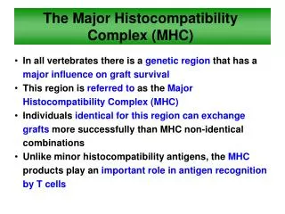
Role of MHC in immune response • TCR recognizes Ag presented in MHC • Context is important • Binding of Ag peptides in non-covalent • Two types of MHC (class I and class II) are recognized by different subsets of T cells • CTL recognizes Ag peptide in MHC class I • T-helper recognizes Ag peptide in MHC class II
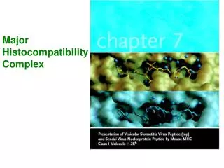
Structure of MHC class I • Two polypeptide chains • Long α chain and short β
Structure of MHC class I • Four regions • Cytoplasmic contains sites for phosphorylation and binding to cytoskeleton • Transmembrane contains hydrophobic AAs • Highly conserved α3 domain binds CD8 • Highly polymorphic peptide binding region formed by α1 and α2
Structure of MHC class I Ag-binding groove • Groove composed of • α helix on 2 opposite walls • Eight β sheets as floor • Residues lining floor are most polymorphic • Groove binds peptides 8-10 AA long
Structure of MHC class I Ag-binding groove • Specific amino acids on peptide are required for “anchor site” in the groove • Many peptides can bind • Interactions at N and C-terminus are critical and “lock” peptide in grove • Center of peptide bulges out for presentation • Consideration in vaccine development
Structure of MHC class II • Two polypeptide chains • α and β • approx equal length
Structure of MHC class II • Four regions • Cytoplasmic contains sites for phosphorylation and binding to cytoskeleton • Transmembrane contains hydrophobic AAs • Highly conserved α2 and β2 domains binds CD4 • Highly polymorphic peptide binding region formed by α1 and β1
Structure of MHC class II Ag-binding groove • Groove composed of • α helix on 2 opposite walls • Eight β sheets as floor • Both α1 and β1 make up groove • Residues lining floor are most polymorphic • Groove binds peptides 13-25 AA long (some outside groove)
Important aspects of MHC • Individuals have a limited number of MHC alleles for each class • High polymorphism in MHC for a species • Alleles for MHC genes are co-dominant • Each MHC gene product is expressed on surface of individual cell
Important aspects of MHC • Each MHC has ONE peptide binding site • But each MHC can bind many different peptides • Only one at a time • Peptide binding is “degenerate” • MHC polymorphism is determined in germline • NO recombination mechanisms for creating diversity in MHC • Peptide must bind with individual’s MHC to induce immune response
Important aspects of MHC • How do peptides get into MHC groove? • Class I: peptides in cytosol associate with MHC • Class II: peptides from within vesicles associate with MHC Peptide in vesicle Displaces Ii chain golgi Cytoplasmic peptide Ii chain ER Class I Class II
Important aspects of MHC • MHC molecules are membrane-bound • Recognition by Ts requires cell-cell contact • Mature Ts must have TCR that recognizes particular MHC • Cytokines (especially IFN-γ) increase expression of MHC
Role of TCR in immune response • Surface molecule on Ts • Recognize Ag presented in MHC context • Similar to Immunoglobulin • Two types of TCR • αβ: predominant in lymphoid tissues • γδ: enriched at mucosal surfaces
Structure of the TCR (αβ) • Heterodimer • α and β chains • approx equal length
Structure of the TCR (αβ) • Regions • Short cytoplasmic tail- cannot transduce activation signal • Transmembrane with hydrophobic AAs • Both α and β have a variable (V) and constant (C) region • V region is hypervariable, determines Ag specificity
Important aspects of TCR • Each T cell has TCR of only ONE specificity • Allelic exclusion • αβ TCR recognizes Ag only in the context of cell-cell interaction and in correct MHC context • γδ TCR recognizes Ag in MHC-independent manner • Response to certain viral and bacterial Ag
Genetic basis for receptor generation • Accomplished by recombination of V, D and J gene segments • TCR β chain genes have V, D, and J • TCR α chain genes have V and J
TCR and CD3 complex • TCR is closely associated with CD3 complex • Group of 5 proteins • Commonly called “invariant” chains of TCR • Role of CD3 complex • CD3 necessary for cell surface expression of TCR • transduces signal after Ag interaction with TCR
The “immunological synapse” • TCR-MHC interaction is not strong • Accessory molecules stabilize interaction • CD4/MHC class II or CD8/MHC class I • CD2/LFA-3 • LFA-1/ICAM-1
The “immunological synapse” • Specificity for Ag is solely in TCR • Accessory molecules are invariant • Cytokines change expression levels
The “immunological synapse” • Co-stimulation is also necessary for activation of T cells • CD28/CD80 or CD86 • CTLA-4 on T cells can also ligate CD80/CD86 • Inhibitory signal • downregulation
Key steps in T cell activation • APC must process and present peptides to Ts • Ts must receive co-stimulatory signal • Accessory adhesion molecules stabilize binding of TCR and MHC • Signal from cell surface is transmitted to nucleus • Cytokines produced help drive cell proliferation