Download
1 / 9
100 likes | 284 Views
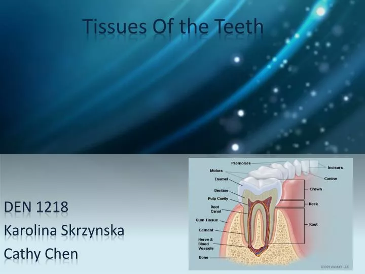
Tissues Of the Teeth. DEN 1218 Karolina Skrzynska Cathy Chen. Tooth Crown. This is the part of the tooth that we see in the mouth It is made up of the enamel, dentin and pulp The crown and the root meet at the neck of the tooth. Crown. Roots. Enamel.

E N D
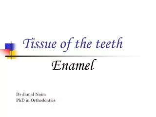
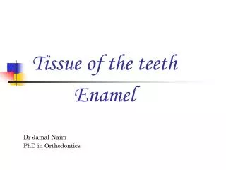
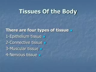
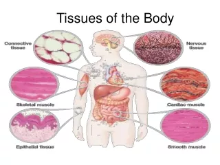
Tissues Of the Teeth DEN 1218 Karolina Skrzynska Cathy Chen
Tooth Crown • This is the part of the tooth that we see in the mouth • It is made up of the enamel, dentin and pulp • The crown and the root meet at the neck of the tooth Crown Roots
Enamel • The enamel is the white hard covering over the crown of the tooth • It is the hardest material in the body and does not have a nerve supply • It is shaped into cusps, fissures and pits in premolar and molar teeth • Enamel consists of approximately 96% of inorganic material and 4% organic material • Enamel's primary mineral is hydroxyapatite • The basic unit of enamel is called an enamel rod
Dentin • Supports the enamel on teeth • It’s a yellow bone like material that’s softer than enamel • It is covered by enamel on the crown and by cementum on the roots • Dentin surrounds and protects the nerves and blood vessels in the crown • Unlike enamel, dentin continues to form throughout life and can be initiated in response to stimuli
Pulp • The nerves and blood vessels of the tooth are called the pulp • The pulp occupies the root canals and the pulp chambers in the crown of the tooth • The pulp provides nutritive, sensory, protective and formative functions • When exposed to infection by decay or injury it will die and cause severe pain
Gingiva Gingiva are part of the soft tissue lining of the mouth. They surround the teeth and provide a seal around them. The gingiva is divided anatomically into--- • Marginal: forms the soft tissue wall of the gingival sulcus. It is supported and stabilized by the gingival fibers. • Attached: is firm, resilient, and tightly bound to the underlying periosteum of alveolar bone • Interdental:occupies the gingival embrasure (the interproximal space beneath the area of tooth contact)
Cementum Cementumis a specialized calcified substance covering the root of the tooth • It is formed continuously throughout life because a new layer of cementum is deposited to keep the attachment intact as the superficial layer of cementum ages.
Periodontal Ligament Types of fibers • Transseptal fibers: extend interproximally over the alveolar bone crest, and form an interdental ligament. • Alveolar crest fibers: extend obliquely from the cementum beneath the junctional epithelium to the alveolar crest. They prevent the extrusion of the tooth and resist lateral tooth movements • Horizontal fibers: attach to the cementum apical to the alveolar crest fibers and run perpendicularly from the root of the tooth to the alveolar bone. • Oblique fibers: are the most numerous fibers , running from cementum in an oblique direction to insert into bone coronally • Apical fibers: extend from cementum around the apex of the root to the bone, forming base of the socket • Interradicular fibers: only found between the roots of multi-rooted teeth, attach from the cementum and insert to the nearby alveolar bone PDL is a group of specialized connective tissue fibers that essentially attach a tooth to the alveolar bone
Alveolar Bone • It contains a region of compact bone adjacent to the periodontal ligament • On the maxilla, the alveolar process is a ridge on the inferior surface • On the mandible, it is a ridge on the superior surface.