Download
1 / 1
10 likes | 109 Views
Supplemental Figure 1. Alignment of CALTPI and CALTPII proteins residues with tobacco lipid transfer protein1 (accession no. D13952).

E N D
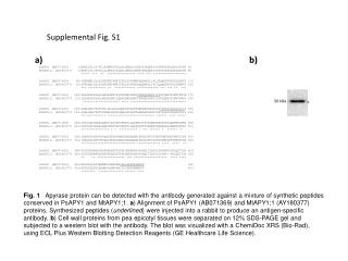
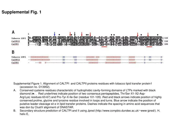
Supplemental Figure 1. Alignment of CALTPI and CALTPII proteins residues with tobacco lipid transfer protein1 (accession no. D13952). • Conserved cysteine residues characteristic of hydrophobic cavity-forming domains of LTPs marked with black diamond (♦). Red underlines indicate position of two consensus pentapeptides, Thr/Ser-X1-X2-Asp-Arg/Lys( residues 63-67) and Pro-Tyr-X-Ile-Ser (residue 101-105) .Red and black arrows indicate position of highly conserved proline, glycine and tyrosine residue involved in loops and turns. Blue arrow indicate the position of putative leader cleavage sit e in lipid transfer proteins. Dashes indicate the spacing in amino acid sequences that was don by ClustV alignment of DNASTAR. • Secondary structure prediction of CALTPI and II using Jpred (http://www.compbio.dundee.ac.uk/~www-jpred/). H, helix E, Supplemental Fig. 1