Download
1 / 17
170 likes | 297 Views
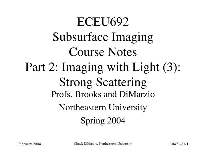
ECEU692 Subsurface Imaging Course Notes Part 2: Imaging with Light (3): Strong Scattering. Profs. Brooks and DiMarzio Northeastern University Spring 2004. Topic Outline. Our Old Scattering Model What We Left Out Phase Functions Multiple Scattering Computational Approaches Reduced Albedo

E N D
ECEU692Subsurface ImagingCourse NotesPart 2: Imaging with Light (3):Strong Scattering Profs. Brooks and DiMarzio Northeastern University Spring 2004 Chuck DiMarzio, Northeastern University
Topic Outline • Our Old Scattering Model • What We Left Out • Phase Functions • Multiple Scattering • Computational Approaches • Reduced Albedo • Photon Migration • Monte-Carlo • A Simple 2-Layer Model Chuck DiMarzio, Northeastern University
Recall Weak Scattering: What is Wrong? Chuck DiMarzio, Northeastern University
Imaging and Multiple Scattering Multi-View Tomography; (e.g. Diffusive Optical Tomography) Localized Probing; (e.g. Confocal microscopy, Optical Coherence Tomography, Two-photon microscopy) Chuck DiMarzio, Northeastern University
Single Scattering Phase Function 10057p2-1 here Chuck DiMarzio, Northeastern University
Multiple Scattering (1) • Scattering Angles Add • Think in 2 Dimensions • Total isSqi • PDF is Convolution of Individual PDF’s Chuck DiMarzio, Northeastern University
Multiple Scattering (2) • Computational Approaches • Reduced Albedo • Photon Migration • Monte-Carlo • Issues • Multiple Scattering • Internal Reflection • Integrated Fresnel Put 10057p2-2 here Chuck DiMarzio, Northeastern University
Assumptions Radiative Transport Equation Taylor Series for Radiance Boundary Conditions Result Diffusion-Like Equation for Photon Density Solution Photon Density Wave DC or AC Complex k (Lossy Wave) Details Another Day Photon Migration Chuck DiMarzio, Northeastern University
W’, Transport Albedo Reflection vs. Albedo: Example Patterson, Michael S., Ephraim Schwartz, and Brian C. Wilson, “Quantitative Reflectance Spectrophotometry for the Noninvasive Measurment of Photosensitizer Concentration in Tissue During Photodynamic Therapy,” Photodynamic Therapy Mechanisms, SPIE Volume 1065, 1989. Pp. 115--122. Chuck DiMarzio, Northeastern University
Loop on Photons Launch Loop on Particle Random Distance Scatter or Absorb Random Angle Exit - Save Data Compute Statistics Widely Applicable Accurate Phase Function not Needed? Computationally Intensive Monte-Carlo Models Chuck DiMarzio, Northeastern University
1c. Scatter or Absorb 1a. Launch 1d. Random Direction 1b. Random Distance 2-N. Next Photons 1e. Continue or Exit: If Exit, count as Reflection or Transmission N+1. Summarize Statistics Refl Radius Monte-Carlo Operation Chuck DiMarzio, Northeastern University
Simple Skin Model R sc • Rsc and Re • No desired signal • Rd < Rd (no blood), • Rd contains wanted signal • Neglecting • index of refraction changes • multiple scattering between layers • specular reflections. R R ( l ) e d Stratum Corneum 100 m m Epidermis Arterial Hb 2 mm Dermis Venous Hb Thanks to Peter Dwyer at Lucid Technologies and MGH Chuck DiMarzio, Northeastern University
Optical Properties of Human Tissue Scattering Absorption Epidermis HbO2 Dermis, Hb, HbO2 Melanin Hb Epidermis Anisotropy Dermis Hb, HbO2 Dermis Water Epidermis Chuck DiMarzio, Northeastern University
No Hb HbO2 Hb Sample Monte-Carlo-Model Spectra for Tissue • Inputs: • Epidermis = 0.065 mm • Dermal Hb = 0.12 mM • Outputs: • Reflectance (Shown) • Radially Resolved • Reflectance • Angular Resolved • Reflectance Chuck DiMarzio, Northeastern University
Simplified Skin Model Equations R = Rsc + Re + Rd R = Rsc + Re + Rd + Hb Spectral Signatures Chuck DiMarzio, Northeastern University
Simplified Skin Model Equations (Cont.) • Want to find s = oxygen saturation of Hb • ma depends on s ma = koxycs + kdeoxyc(1-s) + ma0 koxy = Molar absorption coefficient of oxy-Hb kdeoxy = Molar absorption coefficient of deoxy-Hb c = Concentration of Hb s = Oxygen saturation fraction ma0 = Other chromophores besides blood • Approximation: wavelengths close together only ma depends on l Chuck DiMarzio, Northeastern University
(BC) A R0 s Simplified Skin Model Equations (Cont.) • Grouping some variables together, to solve for s • Four equations, four unknowns Chuck DiMarzio, Northeastern University