Download
1 / 52
540 likes | 1.08k Views
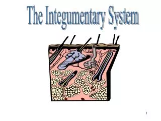
Integumentary System & Wound Symposium. Wound Debridement. Significance of Necrotic Tissue. As tissues die, they change in color, consistency, and adherence to the wound bed. As NT increases in severity color changes from White/Grey to Tan or Yellow and finally to Brown or Black

E N D
Integumentary System & Wound Symposium Wound Debridement
Significance of Necrotic Tissue • As tissues die, they change in color, consistency, and adherence to the wound bed. • As NT increases in severity color changes from White/Grey to Tan or Yellow and finally to Brown or Black • Consistency changes as tissues dessicate or dry • Eventually NT becomes dry leathery and hard
Significance of Necrotic Tissue • Wound etiology influence clinical appearance • Subcutaneous fat forms stringy, yellow slough • Muscle Tissue degenerates into thick, tenacious tissue • Hard Black Eschar = Full-Thickness destruction • Grey/Blueness or white devitalized tissue may represent prolonged ischemia
Slough • Yellow (or) Tan • Thin, mucinous or stringy Sussman, C., Bates Jensen, B. (2001). Wound Care 2nd addition. Aspen, Gaithersberg, Md. Sussman, C., Bates Jensen, B. (2001). Wound Care 2nd addition. Aspen, Gaithersberg, Md.
Eschar • Brown or Black • Soft or Hard • Full-thickness destruction • ** The more water content present, the less adherent the debris is to the wound bed.
Sussman, C., Bates Jensen, B. (2001). Wound Care 2nd addition. Aspen, Gaithersberg, Md.
Adherence • Adhesiveness of debris • Ease at which the two are separated • NT becomes more adherent to the wound as level of damage increases • Eschar more adherent than yellow slough
Necrotic Tissue • Retards Wound Healing • Medium for Bacterial Growth • Physical Barrier to Epidermal Resurfacing, Contraction & Granulation • More NT = More Healing Time • NT Obscures Visualization of the Total Wound
Arterial/Ischemic Wounds • NT may appear as dry gangrene • Thick, dry, dessicated, black/gray appearance • Firmly adhered to wound bed • May be surrounded with a red halo Sussman, C., Bates Jensen, B. (2001). Wound Care 2nd addition. Aspen, Gaithersberg, Md
Neurotrophic Wounds • Usually no necrosis • Often have hyperkeratosis surrounding the wound • Hyperkeratosis looks like callus formation at the wound edges (From: Myers, B.A. (2004).Wound Management Principles and Practice. Prentice Hall, Saddle River, NJ)
Venous Disease Wounds • Either Eschar or Slough • Yellow fibrinous material covers the wound • Eschar might be due to dessication and or necrotic debris
Pressure Sores • NT relates to amount of tissue destruction • Early stage of pressure ulcer, tissue may appear hard (indurated)with purple or black discoloration on intact skin (indicative of tissue death) Fitzpatrick, T.B., Johnson, R.A., Wolff, K., Polano, M.K., Suurmond D. (1997). Color Atlas and Synopsis of Clinical Dermatology: Common and Serious Diseases. McGraw-Hill: Health Professions Division: New York.
Intervention: Debridement • Prevent bacteria from colonizing • Prevent competition with viable cells for oxygen and nutrients • Removal of necrotic and/or infected tissues that interfere with wound healing • Debridement & Irrigation are reported to be the most effective method of controlling wound colonization
Appropriate Wounds for Debridement • Partial or Full-thickness wounds • Clinical Signs of Inflammation or Infection: • Periwound erythema • Warmth • Induration • Edema • Foul Odor • Non-viable tissue or purulent exudate
Clinical Considerations • Viable wound and periwound tissues are adequately perfused with blood • Precautions relative to introducing pathogens must be observed • Debridement of dry eschar over a bone or tendon is contraindicated • Debridement is contraindicated in the presence of dry gangrene • Caution must be exercised when debriding a wound of a patient on anticoagulants
Debridement • Improves wound and soft tissue status • Reduces risk of infection, complications, and secondary impairments • Enhances Wound Healing • With Debridement Wounds get “Bigger Before They Get Better”
Identification of Tissue Types • Skin • Epidermis:outer avascular layer • 0.06 -.6mm thick, sloughs Q 30 days • Waterproof keratinocytes are located in the epidermis • New cells located in the basal layer
Identification of Tissue Types • Skin • Basement Membrane • Dermal-epidermal junction • Separates and attaches the epidermis and the dermis • Atrophies with aging (skin tears)
Identification of Tissue Types • Skin • Dermis • Provides support and nutrition for the epidermis • Fibroblasts produce collagen for tensile strength • Fibroblasts synthesize elastin for resiliency, produce other components of ground substance (GAGS, Proteoglycans, glycoproteins) • Hair follicles, sweat glands, nails, blood vessels and nerves located in the dermis.
Identification of Tissue Types • Subcutaneous Layer • Insulation • Nutrition • Cushioning • Composed of: • adipose tissue • Major vessels • Lymphatics • Nerves • Easily Damaged by Pressure & Infection
Identification of Tissue Types • Fascia • Shiny white & surrounds skeletal muscle • Infection (e.g., necrotizing fasciitis) is spread easily along facial planes • Precaution: • When fascial planes are separated or penetrated the risk of bacterial invasion increase.
Identification of Tissue Types • Skeletal Muscle • Purpose is to provide function • Protects: bones, joints, nerves, and vessels • Pads bony prominences • Healthy muscle is dull red, contractile, and vascular • Necrotic muscle is a darker dull red and avascular
Identification of Tissue Types • Bone • If healthy, hard & white • Cortical bone covered with periosteum • Periosteum is richly vascularized • Provides surface for granulation tissue formation • Accepts skin graft if healthy • If exposed it will dessicate, & turn yellow and will not allow for granulation • If exposed, cortical bone must be kept moist or it will become necrotic
Identification of Tissue Types • Cartilage • Connective tissue that covers and cushions the articular surface of bone at a joint. • Poor vascularity
Identification of Tissue Types • Blood Vessels • Arteries • Arterioles • Capillaries • Venules • Veins • Understanding Anatomy is crucial to avoid damage
Identification of Tissue Types • Tendon • Strong, elastic, fibrous tissues • Attach muscles to bones • When exposed can be identified by manually moving the adjacent joint • Poor vascularity, become infected easily • Must be kept moist if exposed • Healthy tendons are shiny white and are covered with paratenon • Paratenon carry blood, should not be debrided if healthy as the tendon will become necrotic without it. • A necrotic tendon will not become viable again • Loss of function results from loss of tendon
Sussman, C., Bates Jensen, B. (2001). Wound Care 2nd addition. Aspen, Gaithersberg, Md
Sussman, C., Bates Jensen, B. (2001). Wound Care 2nd addition. Aspen, Gaithersberg, Md
Debridement • Removal of necrotic & extraneous (foreign material, debris) tissue from a wound
Purpose of Debridement • Decrease bacteria within the wound bed, decreasing risk of infection • Increase the effect of topical antimicrobials • Improve the effect of inflammatory cells • Decrease the length of the inflammatory phase • Decrease the metabolic expense for healing • Decrease the physical barrier to healing • Decrease odor of the wound
Types of Debridement • Non-Selective • Selective
Non-Selective • Mechanical Debridement • Dry to Dry • Wet to Dry • Wet to Wet • Dakin’s Solution • Hydrogen Peroxide • W/P • Irrigation/ Lavage
Selective • Autolytic: Use of body’s own endogenous enzymes. Apply a moisture retentive dressing/Saran Wrap. Wound fluid trapped beneath the dressing softens & liquefies necrotic tissue. Growth factors and inflammatory cells may enhance healing as well. • Least invasive, least painful, consist with moist wound healing model • Contraindicated in infected wounds • Enzymatic: Use of topical exogenous enzymes to remove devitalized tissue • Elase, Santyl, Accuzyme, Panafil • Sharp/Surgical: Scalpel, Forceps, Scissors
Autolytic Debridement Protocol (From: Myers, B.A. (2004).Wound Management: Principles and Practice. Prentice Hall, Saddle River, NJ)
Enzymatic Debridement • Indicated for infected & uninfected wounds with necrotic tissue • In infected wounds, enzymes may be used with topical antimicrobial therapy (ex. Polymoxin B with Collagenase) • Contraindicated • Wounds with exposed deep tissues (ligament, tendon, capsule, bone, nerve, muscle, blood vessels) • Discontinue after 2 weeks if NT is not effectively reduced
Enzymatic Debridement Protocol (From: Myers, B.A. (2004).Wound Management: Principles and Practice. Prentice Hall, Saddle River, NJ)
Talking Points • Dry Eschar • Cross Hatch or put dressing on to rehydrate • Enzymes are tough to activate, do better in moist environment From: Myers, B.A. (2004).Wound Management: Principles and Practice. Prentice Hall, Saddle River, NJ)
Sharp Debridement • Removal of nonviable tissue with sterile instruments • Physicians, Nurses, PA & PT’s • No State Practice Act denies PT’s the right to perform Sharp Debridement • All PT Practice Acts are written broadly enough to allow PT’s to perform wound debridement without restriction • Arkansas, Arizonia, California, Colorado, Hawaii, Montana, New Hamshire, New Mexico, Nortyh Carolina, South Carolina, Tennessee, Texas and Utah specifically cite wound debridement in their PT Practice Acts • PTA’s cannot perform sharp debridement
Indications for Sharp Debridement • Large Amount of Necrotic Tissue • Advancing Cellulitis or Sepsis • Thick Adherent Eschar
Red Yellow Black Color Code • Red • Pale pink to beefy red, granulation tissue • Goals: Protect wound, Maintain warm moist environment, Protect periwound • Yellow • Moist Yellow Slough, may vary in adherence • Goals: Debride necrotic tissue, Absorb drainage, Protect Peri-wound • Black • Thick, Black, adherent eschar • Goals: Debride necrotic tissue Cuzzell, J.Z. Am J. of Nursing (1988)
Debride Necrotic Tissue Eschar, Slough Foreign Material Debris Residual Topical agents Blisters Callus Do not debride Granular Tissue Viable tissue Stable heel ulcer Gangrene, osteo Electrical Burns Deeper Tissues Muscle, tendon, ligament, bone, nerves, blood vessels Avoid in patients with impaired clotting mechanisms Indication & Contraindications
Sharp Debridement • Two Types • Serial Instrumental Debridement • Selective Sharp Debridement
Serial Instrumental Debridement • Uses • Forceps and scissors • Occurs over several visits • Creates minimal bleeding • Usually requires softening necrotic tissue, making it more amenable to debridement, by use of W/P, Irrigation, or Pulsatile Lavage • Goal: Remove loosely adherent necrotic tissue
Selective Sharp Debridement • Uses Scissors and/or Scalpel • Cut along the border of viable & non-viable tissue • Usually, does not require prior tissue preparation • Gelfoam or silver nitrate may be needed to control minimal bleeding • Requires use of dry dressing for 8-24 hrs after debridement
Contraindications to Sharp Debridement • When area of debridement cannot be adequately visualized (tunneling or undermining) • When material to be debrided cannot be identified • When clinician is out of her or his comfort zone • When competency has not been met • Sharp debridement should not be performed on uninfected ischemic ulcers with low ABI • Only physicians should sharp debride hypergranulated tissue
Sharp Debridement Protocol (From: Myers, B.A. (2004).Wound Management: Principles and Practice. Prentice Hall, Saddle River, NJ)
Use of Instruments: Forceps are used to lift devitalized tissue. Hold the scissors parallel(level) to the specimen to avoid piercing the specimen with the sharp end of the scissors (From: Myers, B.A. (2004).Wound Management: Principles and Practice. Prentice Hall, Saddle River, NJ)
Use of Scalpel: Hold blade level (parallel) with tissue to be debrided Debride in layers to prevent incising healthy tissue Use forceps to apply gentle traction to the devitalized tissue (From: Myers, B.A. (2004).Wound Management: Principles and Practice. Prentice Hall, Saddle River, NJ)
Termination of Sharp Debridement • Clinician becomes fatigued • Patient reports Increased Pain • Patient is less tolerant to procedure • Bleeding beyond minimal • A new facial plane is identified • All necrotic tissue has been removed
Debridement Competency (From: Myers, B.A. (2004).Wound Management: Principles and Practice. Prentice Hall, Saddle River, NJ)