Download
1 / 92
970 likes | 1.3k Views
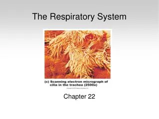
Chapter 22 Respiratory System. Respiration ventilation of lungs exchange of gases between air and blood blood and tissue fluid use of O 2 in cellular metabolism. Organs of Respiratory System. Nose, pharynx, larynx, trachea, bronchi, lungs. General Aspects of Respiratory System.

E N D
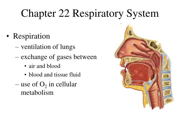
Chapter 22 Respiratory System • Respiration • ventilation of lungs • exchange of gases between • air and blood • blood and tissue fluid • use of O2 in cellular metabolism
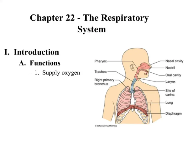
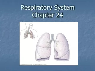
Organs of Respiratory System • Nose, pharynx, larynx, trachea, bronchi, lungs
General Aspects of Respiratory System • Airflow in lungs • bronchi bronchioles alveoli • Conducting division • passages serve only for airflow, nostrils to bronchioles • Respiratory division • alveoli and distal gas-exchange regions • Upper respiratory tract • organs in head and neck, nose through larynx • Lower respiratory tract • organs of the thorax, trachea through lungs
Nose • Functions • warms, cleanses, humidifies inhaled air • detects odors • resonating chamber that amplifies the voice • Bony and cartilaginous supports (fig. 22.2) • superior half: nasal bones medially + maxillae laterally • inferior half: lateral and alar cartilages • ala nasi: flared portion shaped by dense CT, forms lateral wall of each nostril
Nasal Cavity • Extends from nostrils to choanae (posterior nares) • ethmoid and sphenoid bones compose the roof • palate forms the floor • Vestibule: dilated chamber inside ala nasi • stratified squamous epithelium, vibrissae (guard hairs) • Nasal septum divides cavity into right and left chambers called nasal fossae • inferior part formed by vomer • superior part by perpendicular plate of ethmoid bone • anterior part by septal cartilage
Nasal Cavity - Conchae and Meatuses • Superior, middle and inferior nasal conchae • 3 folds of tissue on lateral wall of nasal fossa • mucous membranes supported by thin scroll-like turbinate bones • Meatuses • narrow air passage beneath each conchae • narrowness and turbulence ensures air contacts mucous membranes
Nasal Cavity - Mucosa • Olfactory mucosa lines roof of nasal fossa • Respiratory mucosa lines rest of nasal cavity with ciliated pseudostratified epithelium • Defensive role of mucosa • mucus (from goblet cells) traps inhaled particles • bacteria destroyed by lysozyme
Nasal Cavity - Cilia and Erectile Tissue • Function of cilia of respiratory epithelium • drive debris-laden mucus into pharynx to be swallowed • Erectile tissue of inferior concha • venous plexus that rhythmically engorges with blood and shifts flow of air from one side of fossa to the other once or twice an hour to prevent drying • Spontaneous epistaxis (nosebleed) • most common site is the inferior concha
Pharynx • Nasopharynx (pseudostratified epithelium) • posterior to choanae, dorsal to soft palate • receives auditory tubes and contains pharyngeal tonsil • air turns 90 downward trapping large particles (>10m) • Oropharynx (stratified squamous epithelium) • space between soft palate and root of tongue, inferiorly as far as hyoid bone, contains palatine and lingual tonsils • Laryngopharynx (stratified squamous epithelium) • hyoid bone to cricoid cartilage (inferior end of larynx)
Larynx • Glottis - superior opening • Epiglottis - flap of tissue that guards glottis, directs food and drink to esophagus • Infant larynx • higher in throat, forms a continuous airway from nasal cavity that allows breathing while swallowing • by age 2, more muscular tongue, forces larynx down
Nine Cartilages of Larynx • Epiglottic cartilage • Thyroid cartilage - largest, has laryngeal prominence • Cricoid cartilage - connects larynx to trachea • Arytenoid cartilages (2) - posterior to thyroid cartilage • Corniculatecartilages (2) - attached to arytenoid cartilages like a pair of little horns • Cuneiformcartilages (2) - support soft tissue between arytenoids and the epiglottis
Walls of Larynx • Interior wall has 2 folds on each side, from thyroid to arytenoid cartilages • vestibular folds: superior pair, close glottis during swallowing • vocal cords:produce sound • Intrinsic muscles - rotate corniculate and arytenoid cartilages, which adducts (tightens: high pitch sound) or abducts (loosens: low pitch sound) vocal cords • Extrinsic muscles - connect larynx to hyoid bone, elevate larynx during swallowing
Trachea • Rigid tube 4.5 in. long and 2.5 in. in diameter, anterior to esophagus • Supported by 16 to 20 C-shaped cartilaginous rings • opening in rings faces posteriorly towards esophagus • trachealis spans opening in rings, adjusts airflow by expanding or contracting • Larynx and trachea lined with ciliated pseudostratified epithelium which functions as mucociliary escalator
Bronchial Tree • Primary bronchi (C-shaped rings) • arise from trachea, after 2-3 cm enter hilum of lungs • right bronchus slightly wider and more vertical (aspiration) • Secondary (lobar) bronchi (overlapping plates) • branches into one secondary bronchus for each lobe • Tertiary (segmental) bronchi (overlapping plates) • 10 right, 8 left • bronchopulmonary segment: portion of lung supplied by each
Bronchial Tree 2 • Bronchioles (lack cartilage) • have layer of smooth muscle • pulmonary lobule: portion ventilated by one bronchiole • divides into 50 - 80 terminal bronchioles • terminal bronchioles • have cilia , give off 2 or more respiratory bronchioles • respiratory bronchioles • divide into 2-10 alveolar ducts • Alveolar ducts - end in alveolar sacs • Alveoli - bud from respiratory bronchioles, alveolar ducts and alveolar sacs
Pleurae and Pleural Fluid • Visceral and parietal layers • Pleural cavity and fluid • Functions • reduction of friction • creation of pressure gradient • lower pressure assists in inflation of lungs • compartmentalization • prevents spread of infection
Pressure and Flow • Atmospheric pressure drives respiration • 1 atmosphere (atm) = 760 mmHg • Intrapulmonary pressure and lung volume • pressure is inversely proportional to volume • for a given amount of gas, as volume , pressure and as volume , pressure • Pressure gradients • difference between atmospheric and intrapulmonary pressure • created by changes in volume of thoracic cavity
Inspiration - Muscles Involved • Diaphragm (dome shaped) • contraction flattens diaphragm • Scalenes • fix first pair of ribs • External intercostals • elevate 2 - 12 pairs • Pectoralis minor, sternocleidomastoid and erector spinae muscles • used in deep inspiration
Inspiration - Pressure Changes • intrapleural pressure • as volume of thoracic cavity ,visceral pleura clings to parietal pleura • intrapulmonary pressure • lungs expand with the visceral pleura • Transpulmonary pressure • intrapleural minus intrapulmonary pressure (not all pressure change in the pleural cavity is transferred to the lungs) • Inflation of lungs aided by warming of inhaled air • A quiet breathe flows 500 ml of air through lungs
Passive Expiration • During quiet breathing, expiration achieved by elasticity of lungs and thoracic cage • As volume of thoracic cavity , intrapulmonary pressure and air is expelled • After inspiration, phrenic nerves continue to stimulate diaphragm to produce a braking action to elastic recoil
Forced Expiration • Internal intercostal muscles • depress the ribs • Contract abdominal muscles • intra-abdominal pressure forces diaphragm upward, pressure on thoracic cavity
Pneumothorax • Presence of air in pleural cavity • loss of negative intrapleural pressure allows lungs to recoil and collapse • Collapse of lung (or part of lung) is called atelectasis
Resistance to Airflow • Pulmonary compliance • distensibility of the lungs; the change in lung volume relative to a given change in transpulmonary pressure • decreased in diseases with pulmonary fibrosis (TB) • Bronchiolar diameter • primary control over resistance to airflow • bronchoconstriction • triggered by airborne irritants, cold air, parasympathetic stimulation, histamine • bronchodilation • sympathetic nerves, epinephrine
Alveolar Surface Tension • Thin film of water necessary for gas exchange • creates surface tension that acts to collapse alveoli and distal bronchioles • Pulmonary surfactant (great alveolar cells) • disrupts hydrogen bonds of water, surface tension • As passages contract during expiration, surface tension naturally and surfactant concentration preventing alveolar collapse • Respiratory distress syndrome of premature infants
Alveolar Ventilation • Dead air • fills conducting division of airway, cannot exchange gases • Anatomic dead space • conducting division of airway • Physiologic dead space • sum of anatomic dead space and any pathological alveolar dead space • Alveolar ventilation rate • air that actually ventilates alveoli X respiratory rate • directly relevant to body’s ability to exchange gases
Nonrespiratory Air Movements • Functions other than alveolar ventilation • flow of blood and lymph from abdominal to thoracic vessels • Variations in ventilation also serve • speaking, yawning, sneezing, coughing • Valsalva maneuver • take a deep breath, hold it and then contract abdominal muscles; increases pressure in the abdominal cavity • to expel urine, feces and to aid in childbirth
Measurements of Ventilation • Spirometer • device a subject breathes into that measures ventilation • Respiratory volumes • tidal volume: air inhaled or exhaled in one quiet breath • inspiratory reserve volume: air in excess of tidal inspiration that can be inhaled with maximum effort • expiratory reserve volume: air in excess of tidal expiration that can be exhaled with maximum effort • residual volume: air remaining in lungs after maximum expiration, keeps alveoli inflated
Respiratory Capacities • Vital capacity • amount of air that an be exhaled with maximum effort after maximum inspiration; assess strength of thoracic muscles and pulmonary function • Inspiratory capacity • maximum amount of air that can be inhaled after a normal tidal expiration • Functional residual capacity • amount of air in lungs after a normal tidal expiration
Respiratory Capacities • Total lung capacity • maximum amount of air lungs can contain • Forced expiratory volume (FEV) • % of vital capacity exhaled/ time • healthy adult - 75 to 85% in 1 sec • Peak flow • maximum speed of exhalation • Minute respiratory volume (MRV) • TV x respiratory rate, at rest 500 x 12 = 6 L/min • maximum: 125 to 170 L/min