Download
1 / 48
480 likes | 927 Views
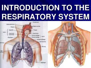
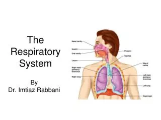
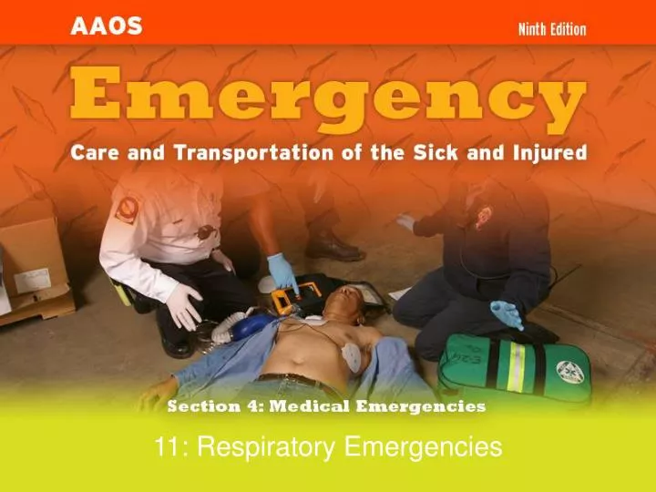
11: Respiratory Emergencies. Respiratory System. Anatomy and Function of the Lung. Characteristics of Adequate Breathing. Normal rate and depth Regular breathing pattern Good breath sounds on both sides of the chest Equal rise and fall of chest Pink, warm, dry skin.

E N D
Characteristics of Adequate Breathing • Normal rate and depth • Regular breathing pattern • Good breath sounds on both sides of the chest • Equal rise and fall of chest • Pink, warm, dry skin
Characteristics ofInadequate Breathing • Pulmonary vessels become obstructed. • Alveoli are damaged. • Air passages are obstructed. • Blood flow to the lungs is obstructed. • Pleural space is filled.
Signs ofInadequate Breathing • Slower than 12 breaths/min or faster than 20 breaths/min • Unequal chest expansion • Decreased breath sounds • Muscle retractions • Pale or cyanotic skin • Cool, damp (clammy) skin • Shallow or irregular respirations • Pursed lips • Nasal flaring
Dyspnea • Shortness of breath or difficulty breathing • Patient may not be alert enough to complain of shortness of breath.
Upper or Lower Airway Infection • Infectious diseases may affect all parts of the airway. • The problem is some form of obstruction to the air flow or the exchange of gases.
Acute Pulmonary Edema • Fluid build-up in the lungs • Signs and symptoms • Dyspnea • Frothy pink sputum • History of chronic congestive heart failure • Recurrence high
Chronic Obstructive Pulmonary Disease (COPD) • COPD is the result of direct lung and airway damage from repeated infections or inhalation of toxic agents. • Bronchitis and emphysema are two common types of COPD. • Abnormal breath sounds may be present. • Rhonchi and wheezes
Asthma • Common but serious disease • Asthma is an acute spasm of the bronchioles. • Wheezing may be audible without a stethoscope.
Spontaneous Pneumothorax • Accumulation of air in the pleural space • Caused by trauma or some medical conditions • Dyspnea and sharp chest pain on one side • Absent or decreased breath sounds on one side
Anaphylactic Reactions • An allergen can trigger an asthma attack. • Asthma and anaphylactic (allergic) reactions can be similar. • Hay fever is a seasonal response to allergens.
Pleural Effusion • Collection of fluid outside lung • Causes dyspnea • Caused by irritation, infection, or cancer • Decreased breath sounds over region of the chest where fluid has moved the lung away from the chest wall • Eased if patient is sitting up
Mechanical Obstruction of the Airway • Be prepared to treat quickly. • Obstruction may result from the position of head, the tongue, aspiration of vomitus, or a foreign body. • Opening the airway with the head tilt-chin lift maneuver may solve the problem.
Pulmonary Embolism • A blood clot that breaks off and circulates through the venous system • Signs and symptoms • Dyspnea • Acute pleuritic pain • Hemoptysis • Cyanosis • Tachypnea • Varying degrees of hypoxia
Hyperventilation • Overbreathing resulting in a decrease in the level of carbon dioxide • Signs and symptoms • Anxiety • Numbness • A sense of dyspnea despite rapid breathing • Dizziness • Tingling in hands and feet
You are the Provider • You and your EMT-B partner are dispatched to 1465 Dalles Military Rd for a 33-year-old woman with difficulty breathing. • You arrive at the office building and an upset man identifies himself as the patient’s coworker. • He tells you that the patient has had breathing problems before, but he’s never seen it this bad.
You are the Provider (continued) • He leads you to a woman who is standing with her arms outstretched on the desk with a metered-dose inhaler in hand. • She acknowledges your presence with a nod. When you ask her what is wrong, she answers with a two-word response, “can’t breathe.” • You hear audible wheezes.
Scene Size-UP • How significant is the person’s response to your question and why? • What should you do next? Should you transport this patient or wait for ALS to arrive on scene?
Initial Assessment • Perform initial assessment. • Place the patient on oxygen. • If patient is in respiratory distress, ventilate. • Check pulse.
Difficulty breathing Altered mental status Anxiety or restlessness Increased or decreased respirations Increased heart rate Irregular breathing Cyanosis Signs and Symptoms (1 of 2)
Pale conjunctivae Abnormal breath sounds Difficulty speaking Use of accessory muscles Coughing Tripod position Barrel chest Signs and Symptoms (2 of 2)
You are the Provider (continued) • You arrange to rendezvous with ALS. • You apply high-flow oxygen and obtain the following vital signs: • Pulse: 42 breaths/min • Pulse oximetry: 90% • The patient indicates that she has used the inhaler twice already.
You are the Provider (continued) • What can you do before you meet ALS? • Another pulse oximetry reading reveals a reading of 72%. • The patient is using accessory muscles to breathe. • What do these signs indicate?
COPD Patients • COPD patients cannot handle pulmonary infections well • Usually age 50 or older • History of recurring lung problems • Long-term smokers • Tightness in chest/constant fatigue
Focused History and Physical Exam • Abnormal breath sounds are symptomatic of COPD • Long history of dyspnea with sudden increase in shortness of breath • Recent chest cold with fever • Vital signs • Normal blood pressure • Rapid, occasionally irregular pulse • Respirations rapid or very slow
Interventions • Treat immediate life threats • Possible interventions • Oxygen via nonrebreathing mask at 15 L/min • Positive pressure ventilations • Airway adjuncts • Positioning • Respiratory medications
Detailed Physical Exam • Performed only once life threats are addressed. • May not be able to do if busy treating airway or breathing problems.
Ongoing Assessment • Carefully watch patients for shortness of breath. • Reassess vital signs. • Ask patient if treatment has made a difference. • Check for accessory muscle use.
Emergency Medical Care • Give supplemental oxygen at 10 to 15 L/min via nonrebreathing mask. • Patients with longstanding COPD may be started on low-flow oxygen (2 L/min). • Assist with inhaler if available. • Consult medical control.
Trade names Proventil Ventolin Alupent Metaprel Brethine Generic names Albuterol Metaproterenol Terbutaline Medications in MDI
Prescribed Inhalers • Actions • Relax the muscles surrounding the bronchioles • Enlarge the airways leading to easier passage of air • Side effects • Increased pulse rate • Nervousness • Muscle tremors
Prior to Administration • Read label carefully. • Verify it has been prescribed by a physician for this patient. • Consult medical control. • Make sure the medication is indicated. • Check for contraindications.
Contraindications for MDI • Patient unable to help coordinate inhalation • Inhaler not prescribed for patient • No permission from medical control • Maximum dose prescribed has been taken.
Administration of MDI (1 of 3) • Obtain order from medical control or local protocol. • Check for right medication, right patient, right route. • Make sure the patient is alert. • Check the expiration date. • Check how many doses have been taken.
Administration of MDI (2 of 3) • Make sure inhaler is at room temperature or warmer. • Shake inhaler. • Stop administration of oxygen. • Ask the patient to exhale deeply and put lips around opening. • If the inhaler has a spacer, use it.
Administration of MDI (3 of 3) • Have the patient depress the inhaler and inhale deeply. • Instruct the patient to hold his or her breath. • Continue administration of oxygen. • Allow the patient to breathe a few times then repeat dose according to protocol.
Reassessment • Carefully watch for shortness of breath. • 5 minutes after administration: • Obtain vital signs again. • Perform focused reassessment. • Transport and continue to assess breathing.
Upper or Lower Airway Infection • Administer warm, humidified oxygen. • Do not attempt to suction the airway or insert an oropharyngeal airway in a patient with suspected epiglottitis. • Transport patient in position of comfort.
Acute Pulmonary Edema • Administer 100% oxygen. • Suction secretions. • Transport in position of comfort.
Chronic Obstructive Pulmonary Disease (COPD) • Assist with prescribed inhaler if patient has one. • Transport promptly in position of comfort.
Spontaneous Pneumothorax • Administer oxygen. • Transport in position of comfort. • Monitor closely.
Asthma • Obtain history. • Assess vital signs. • Assist with inhaler if patient has one. • Administer oxygen. • Transport promptly.
Pleural Effusion • Definitive treatment is performed in a hospital. • Administer oxygen and support measures. • Transport promptly.
Obstruction of the Airway • Clear airway. • Administer oxygen. • Transport promptly.
Pulmonary Embolism • Administer oxygen. • Place patient in comfortable position, usually sitting. • Assist breathing as necessary. • Keep airway clear. • Transport promptly.
Hyperventilation • Complete initial assessment and history of the event. • Assume underlying problems. • Do not have patient breathe into a paper bag. • Give oxygen. • Reassure patient and transport.