Download
1 / 47
470 likes | 592 Views
Effective Practices for Description of Science Content within Digital Talking Books. Before the workshop begins, please take a short survey: ncam.wgbh.org/ncam_workshop_surveys.html. WGBH & Access. The Caption Center (est. 1972) Captions television, home videos, feature films CD & DVD-ROM

E N D
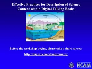
Effective Practices for Description of Science Content within Digital Talking Books Before the workshop begins, please take a short survey: ncam.wgbh.org/ncam_workshop_surveys.html
WGBH & Access The Caption Center (est. 1972) • Captions television, home videos, feature films • CD & DVD-ROM • Streaming video Descriptive Video Service (est. 1990) • Describes television, home videos, feature films by inserting key visual elements during pauses in dialogue National Center for Accessible Media (est. 1993) • supports national policy decisions • develops technical solutions • conducts research • promotes advocacy via outreach
STEM Image Description DAISY Digital Talking Books • Provides standard for accessible digital books NIMAS: National Instructional Materials Accessibility Standard • Requires publishers to provide textbooks in digital form, including images, for students with print disabilities • Without image descriptions, many dtbs remain largely inaccessible. National Science Foundation • Funded NCAM to develop guidelines for making STEM images accessible
Project Partners NCAM American Foundation for the Blind American Printing House for the Blind Recording for the Blind and Dyslexic George Kersher, RFBD and DAISY Larry Scadden, Scientist and frmr NSF Program Officer
Project Goal Provide Research-Based Guidelines for making STEM Images Accessible. Project Process • Collect Images and Descriptions • Evaluate And Refine Descriptions • 2 Round Delphi Survey • 15 Blind STEM Professionals and graduate students • 15 Sighted “Describers” from AFB, RFBD and APH • User Testing • 60 B/VI STEM Focused Adults (including STEM Professionals and Graduate Students) • Publish Results
STEM Description Guidelines ncam.wgbh.org/publications/stemdx
Brevity The most frequent recommendation from respondents was for more brevity in description. It takes people with visual impairments more time to read books and articles than people without visual impairments and the process should not be further slowed down by unnecessarily long image descriptions.
Brevity PREFERRED Descriptive Practice: • The Venn diagram shows 2 intersecting circles, one labeled Africa 93 and the other labeled Asia 155. The area of intersection is labeled 70 PREVIOUS Descriptive Practice: • The figure is a Venn diagram and shows 2 intersecting circles inside a large rectangle. The circles do not touch the rectangle. The circle on the left is labeled Africa and the number 93 is under Africa and above the circle. The circle on the right is labeled Asia and the number 155 is under Asia and above the circle. The intersection of the 2 circles is shaded and has the number 70 in the shaded region.
Data Description should focus on the data and not extraneous visual elements. Elaborately illustrated diagrams often contain key data that can be made accessible by presenting the data separate from description of the overall image.
PREVIOUS Description emphasizes visual PREFERRED Description emphasizes data
Clarity If the reader needs to listen to a description several times because it is poorly written or is presented in a confusing manner, then it is not accessible.
Drill-Down Organization Drill-Down = brief summary followed by extended description and/or specific data. Drill-Down organization allows the reader to either continue reading for more information or stop when they have read all they want.
Drill-Down The figure is a pie chart. Title: Figure 5-2. Distribution of injury deaths by intent: United States, 2003-2004. • Unintentional 67% • Suicide 19% • Homicide 11% • Undetermined 3% • Legal intervention or operations of war less than 1%
Tables Tables, charts and graphs should be presented as tables, not as narrative description. Proper coding (captions, table headers, and table data) provide better access to tables than narrative description. Brief summaries or overviews of the charts should be presented before the tables.
Processes Processes that are presented visually can be converted into nested lists with good results. • Flow Charts • Diagrams • Illustrated Chemical Reactions • And More!
PREVIOUS PREFERRED
Mathematics Math equations should be marked up with MathML and rendered in a way that is preferable to the individual reader.
Narrative Description Many STEM images are best described by linear, narrative description or “traditional” description. Follow the guidelines! • Brevity • Drill-Down Organization • Clarity • Emphasis on Data
Narrative Description • The fish embryo is long, narrow and straight. Its head is small, round, and contains gill arches. A large flap extends to the left, from just below the head to the middle of the embryo. A segmented bony structure runs the length of the embryo on the right. • The reptile embryo is much longer and fatter than the fish embryo, but is curled into a fetal position. Its head is bent forward and is twice as large as that of the fish embryo. The reptile embryo has twice as many gill arches as the fish embryo, but the flap on the left side is only half as long. A segmented bony structure runs the length of the embryo on the right. • The bird embryo is curved more than the fish embryo, but is not as long or as curved as the reptile embryo. The head of the bird embryo is almost as large as that of the reptile embryo, but has fewer gill arches. A flap the same size as that of the reptile embryo extends to the left. A segmented bony structure runs the length of the embryo on the right. Arrows point to the gill arches of all three embryos.
Navigation Control Description presented as text is generally preferred over recorded audio because text readers provide superior navigation control. Properly marked up HTML, especially lists and tables, provides speedy and independent access to data that is unavailable through traditional linear, narrative description.
Four Words to Remember Brevity Data Clarity Control
Diagram of the breathing process. • Inhalation • A muscle at the base the lungs, called the diaphragm, moves downward. • Inside the lungs, pressure decreases and air rushes in. • Ribs move upward and outward. • Volume of the chest cavity increases. • Air flows into the nose and mouth. • Exhalation • Diaphragm moves upward. • Inside the lungs, pressure increases and air moves out. • Ribs move downward and inward. • Volume of chest cavity decreases. • Air flows out through the nose and mouth.
A diagram titled: The Promise of Stem Cell Research. • A petri dish is labeled, Cultured Pluripotent Stem Cells. • Arrows connect the dish of Stem Cells to the following items: • Identify drug targets and test potential therapeutics • Toxicity Testing • Tissues/Cells for Transplantation • Bone marrow for leukemia & chemotherapy • Nerve cells for Parkinsons & Alzhiemer's disease • Heart muscle cells for heart disease • Pancreatic islet cells for diabetes • ? (left blank) • Study cell differentiation • Understanding prevention and treatment of birth defects
An illustration labeled, "Geological unconformities." • The illustration shows a cross-section of a grassy hill, with five horizontal layers. The layers alternate between layers of rock and layers of soil. • In one area, a U-shaped section of mixed rocks and soil cuts down from the surface through four layers. This section is labeled "mixed strata." • The hill slopes down to trees and water. The steep slope is not grassy and the layers are visible. This is labeled "exposed buried strata." • On the other side of the water is a smaller hill with three horizontal layers that match the first three layers of the first hill.
This page contains several captioned illustrations of a woodpecker. 1. The central illustration shows a woodpecker gripping a tree branch with its feet. The woodpecker stands upright on the vertical branch. The woodpecker is yellowish-green with brown and white showing on the edges of its wings and tail feathers. Its breast and head are lighter green, with bright red feathers across the top of its head. Its beak is sharply pointed. 2. A diagram shows a woodpecker's skeleton. Caption: The woodpecker is superbly adapted for chipping away wood and picking out insects from cracks. The beak is very strong with a chisel-like tip, and the neck muscles highly developed for hammering. Even more remarkable are the flexible, elongated hyoid bone which enables the tongue to be greatly protruded, and the complex muscles which push the tongue in and out, stiffen it for spearing insects or move it from side to side to explore the crevices of a rotting log. 3. Diagrams compare a woodpecker's foot to a magpie's foot. The magpie's four toes all point roughly forward, like our fingers, while the woodpecker's toes point in all four directions. Caption: The woodpecker's skeleton shows several special adaptions. The skull is particularly sturdy to withstand the force of the blows as it hammers with its beak; the leg bones are large and strong; the end of the spine curves downwards to enable the tail feathers to act as a prop. One toe of the woodpecker's foot is reversed to give a strong grip 4. A diagram shows a woodpecker's fanning tail feathers. Caption: The feathers in the woodpecker's tail are exceptionally stiff, and the outermost feathers are reduced in size, allowing the tail to act as a prop. 5. On a diagram of a woodpecker's skull, labels point to different bones and muscles. • The tip of the woodpecker's tongue: "Horny barbed tip to spear larger insects." • The outside of the woodpecker's long tongue: "Sheath of skin." • The inside the woodpecker's mouth: "Muscles which can stiffen the tongue or move it from side to side." • Muscles at the top of the woodpecker's neck: "Muscles which pull the tongue in." • The back of the woodpecker's skull: "Muscles which when contracted push the tongue out." • A thin bone that starts near the woodpecker's eye and curves backward around the bird's skull to the base of its tongue: "Hyoid bone."
An illustration shows a cross-section of the human heart. The heart is made up of four chambers, two smaller ones on top (the left and right atrium) and two larger ones below (the left and right ventricle.) A series of arteries and veins carry blood to and from the chambers. Valves separate some of the chambers and blood vessels. The diagram includes the following labels. • right atrium: small upper chamber • superior vena cava: carries blood from above into the right atrium • inferior vena cava: carries blood from below into the right atrium • right pulmonary veins: small blood vessels connected to the right atrium • right ventricle: large lower chamber • tricuspid valve: separates the right atrium and right ventricle • pulmonary artery: carries blood from the right ventricle to the lungs • pulmonary valve: separates the right ventricle and pulmonary artery • left atrium: small upper chamber • left pulmonary veins: small blood vessels connected to the left atrium • left ventricle: large lower chamber • mitral valve: separates the left atrium and left ventricle • aorta: carries blood from the left ventricle to the rest of the body • aortic valve: separates the left ventricle and the aorta
Thank You! • There are many more examples on the Guidelines Website. • We will be adding more examples and training sessions. • Please take a short post-training survey at: ncam.wgbh.org/ncam_workshop_surveys.html
Contact Information Bryan Gould Project Manager WGBH National Center for Accessible Media bryan_gould@wgbh.org ncam.wgbh.org/publications/stemdx