Download
1 / 42
430 likes | 632 Views
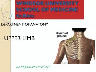
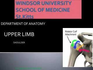
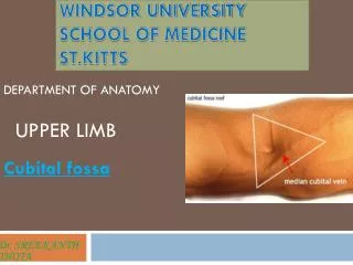
WINDSOR UNIVERSITY SCHOOL OF MEDICINE St.Kitts. DEPARTMENT OF ANATOMY. UPPER LIMB. Dr. SREEKANTH THOTA. Hand . HAND . The hand is the region of the upper limb distal to the wrist joint. It is subdivided into three parts: 1. Wrist 2.Metacarpus

E N D
WINDSOR UNIVERSITYSCHOOL OF MEDICINESt.Kitts DEPARTMENT OF ANATOMY UPPER LIMB Dr. SREEKANTH THOTA Hand
HAND • The hand is the region of the upper limb distal to the wrist joint. • It is subdivided into three parts: • 1. Wrist • 2.Metacarpus • 3.Digits (five fingers including the thumb). • The hand has an anterior surface (palm) and a dorsal surface (dorsum of hand).
Carpal tunnel and structures at the wrist • The carpal tunnel is formed anteriorly at the wrist by a deep arch formed by the carpal bones and the flexor retinaculum.
Carpal arch • The base of the carpal arch is formed medially by the pisiform and the hook of the hamate and laterally by the tubercles of the scaphoid and trapezium.
Flexor retinaculum • flexor retinaculum is a thick connective tissue ligament that bridges the space between the medial and lateral sides of the base of the arch and converts the carpal arch into the carpal tunnel.
Structure and relations • Four tendons of the flexor digitorumprofundus • Four tendons of the flexor digitorumsuperficialis • One tendon of the flexor pollicislongus • Median nerve
Carpal tunnel syndrome • Carpal tunnel syndrome is an entrapment syndrome caused by pressure on the median nerve within the carpal tunnel.
Carpal Tunnel syndrome Common in computer professionals. Due to constant dorsiflexion of wrist while typing the keyboard
Palmaraponeurosis • The palmaraponeurosis is a triangular-shaped condensation of deep fascia that covers the palm and is anchored to the skin in distal regions. • The apex of the triangle is continuous with the palmarislongus tendon.
Dupuytren Contracture of Palmar Fascia • Dupuytren contracture is a disease of the palmar fascia resulting in progressive shortening, thickening, and fibrosis of the palmar fascia and aponeurosis.
Compartments of palm • 1. Hypothenar compartment • 2. Thenar compartment • 3. Central compartment • 4. Adductor compartment • 5.Interosseous compartment
Muscles • The intrinsic muscles of the hand are located in five compartments • All of the intrinsic muscles of the hand are innervated by the deep branch of the ulnarnerve except for the three thenar and two lateral lumbricalmuscles, which are innervated by the median nerve.
Palmaris brevis Action: Improves grip
Adductor pollicis • Action: • Adducts thumb towards middle digit
Movements of thumb • Extension: extensor pollicislongus, extensor pollicisbrevis • Flexion: flexor pollicislongus and flexor pollicisbrevis • Abduction: abductor pollicislongus and abductor pollicisbrevis. • Adduction: adductor pollicis • Opposition: opponenspollicis.
Dorsal interossei Action of Dorsal Interossei : DAB : Abduction Little finger and thumb have no Dorsal interossei muscle
Palmarinterossei Action of Palmarinterossei : PAD :Adduction Middle finger and thumb have no palmarinterossei muscle
Arteries of hand • Blood supply to the hand is by the radial and ulnar arteries, which form two interconnected vascular arches (superficial and deep) in the palm.
Ulnar artery and superficial palmar arch Superficial palmar arch: Ulnar artery+ palmar branch of radial artery
Radial artery and deep palmar arch • Deep palmar arch: Deep palmar branch of ulnar artery+ radial artery
Allen's test • To test for adequate anastomoses between the radial and ulnar arteries, compress both the radial and ulnar arteries at the wrist, then release pressure from one or the other, and determine the filling pattern of the hand.
Nerves • The hand is supplied by the ulnar, median, and radial nerves.
Ulnar nerve • Immediately distal to the pisiform, ulnar nerve divides into a deep branch, which is mainly motor and a superficial branch, which is mainly sensory. Deep branch: supplies the hypothenarinterossei, adductor pollicis, and the two medial lumbricals. Superficial branch: supply skin on the palmar surface of the little finger and the medial half of the ring finger
Ulnar nerve injury • The ulnar nerve is most commonly injured at two sites: • 1. Elbow • 2. wrist • Clawing of the hand: • Metacarpophalangeal joints of the fingers are hyperextendedand the interphalangeal joints are flexed.
Ulnar Canal Syndrome (Guyon Tunnel Syndrome) • Compression of the ulnar nerve may occur at the wrist where it passes between the pisiform and the hook of the hamate.
Median nerve • The median nerve is the most important sensory nerve in the hand because it innervates skin on the thumb, index and middle fingers, and lateral side of the ring finger. • Branches: • 1. Recurrent branch: innervates the three thenar muscles • 2. Palmar digital nerves: In addition to skin, the digital nerves supply the lateral two lumbrical muscles
Ape hand • Refers to a deformity in which thumb movements are limited to flexion and extension of the thumb in the plane of the palm. • Severance of median nerve paralyzes the thenar muscles and the thumb loses much of its usefulness.
Superficial branch of the radial nerve • The only part of the radial nerve that enters the hand is the superficial branch. • Innervates skin over the dorsolateral aspect of the palm and the dorsal aspects of the lateral three and one-half digits distally to approximately the terminal interphalangeal joints.
1 • A 21 year old girl is brought to the emergency department with a puncture wound on the palmar side of her left index finger. Preservation of which of the following movements of her index finger will confirm the functional integrity of the flexor digitorumprofundus muscle? • Flexion of the metacarpophalangeal joint • Adduction • Abduction • Flexion of the distal interphalangeal joint • Flexion of the proximal interphalangeal joint
2. • A 53-year-old African American man involved in a motor vehicle accident sustains a severe mid-shaft fracture of the right humerus. Vitals are Temp-100.0F, BP-120/88mm/Hg, pulse- 118/min, and RR- 14/min. Examination reveals wrist drop and no ulnar or radial pulses in the right arm. Examination reveals decreased sensation over the dorsal aspect of the lateral 3½ digits. The rest of the physical exam is otherwise unremarkable. What nerve is most likely injured given the findings in this patient? • A.Axillarynerve • B.Musculocutaneousnerve • C.Mediannerve • D.Radialnerve • E.Ulnarnerve
3 • The figure below represents cutaneousinnervation of wrist and hand. The area A in the figure represents which Nerve? • Superficial branch of radial nerve • Anterior interosseous nerve • Palmar branch of median nerve • Palmar branch of ulnar nerve • Lateral cutaneous nerve of fore arm