Download
1 / 32
570 likes | 2.02k Views
Glomerulonephritis in children. Pavlyshyn H.A. Definition. Acute glomerulonephritis is the inflammation of the glomeruli which causes the kidneys to malfunction It is also called Acute Nephritis, Glomerulonephritis and Post-Streptococcal Glomerulonephritis

E N D
Glomerulonephritis in children Pavlyshyn H.A.
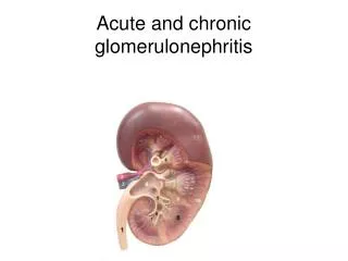
Definition • Acute glomerulonephritis is the inflammation of the glomeruli which causes the kidneys to malfunction • It is also called Acute Nephritis, Glomerulonephritis and Post-Streptococcal Glomerulonephritis • Predominantly affects children from ages 2 to 12 • Incubation period is 2 to 3 weeks
Autoimmune Reactions Some progress as either focal segmental glomerulosclerosis or tubulointerstitial nephritis
Possible Clinical Manifestations Proteinuria – asymptomatic Haematuria – asymptomatic Hypertension Nephrotic syndrome Nephritic syndrome Acute renal failure Rapidly progressive renal failure End stage renal failure 8
Presentation • Hematuria • with Proteinuria • with Dysmorphic rbcs • with Rbc casts • Oliguria • Volume overload • Hypertension
Urine Sediment Analysis G4 cell
Other H&P findings • Neurological changes • Pharyngitis • URI / sinusitis • Hemoptysis • Rash • Murmur • Arthritis • Edema
Membrane attack complex (C4bC2a) (C4 + C2) Recruitment of PMNs C3b C3 C3a Opsonization, phagocytosis C3 convertase Alternative pathway Anaphylaxis, Chemotaxis Microbial surfaces (polysaccharides) Complement Abnormalities Ab-Ag complexes C3 convertase Classical pathway
Differential Diagnosis Hypocomplementemia • PIGN • MPGN • SLE • Cryoglobulinemia • Bacterial Endocarditis • Shunt nephritis Normal complement • HUS • IgAN • HSP • Alport’s / TBMD
b-hemolytic Streptococci • Most common organism in PIGN • 20% children are asymptomatic carriers • Nephritic factor • Host susceptibility factors (HLA-DR) • Treatment of prodromal illness doesn’t prevent nephritis • ASO titers are NOT helpful
Post Infectious GN Pathogenesis • Strep antigens trigger antibodies that cross-react to glomeruli • Circulating immune complexes get filtered by glomerulus & get stuck • Immune complexes activate complement • Diffuse & generalized damage to glomeruli • ↓ GFR due to inflammation, damage to BM • ↓ RBF in proportion to GFR, so filtration fraction normal • Tubular function is preserved • Plasma renin and aldosterone are normal Presentation • 7-14 days after pharyngitis • 14-21 days after impetigo (upto 6 wks) • Abrupt onset
Manifestations of PIGN • Edema 85% • HTN 60-80% • Gross hematuria 25-33% • CNS (i.e. Sz) 10% • Nephrotic syndrome rare • ARF not uncommon • C3 decreased • C4 typically normal
Management of PIGN • Antibiotics do NOT prevent GN • Sodium & Fluid restriction • Antihypertensives, diuretics for HTN • Dialysis if necessary • Prognosis usually excellent • 0.5% mortality due to pulmonary edema or pneumonia • <1% progress to CKD stage 5 • Follow-up • Gross hematuria resolves within 2 weeks • Complement low for 6-8 weeks • Proteinuria remains upto 6 months • Hematuria remains upto 2 years
Histopathology Diffuse = all glomeruli Generalized = all segments of glomeruli
IgG Immunofluorescence Starry Sky Pattern
Basement membrane Foot processes Electron microscopy - Normal
Electron microscopy of PIGN • Subepithelial immune deposits (humps) • Mesangial, subendothelial, intramembranous deposits less common • Effacement of foot processes
Hemolytic Uremic Syndrome • 2 cases/100,000 annually • Peak incidence <5yo (6/100,000) • More common June-September • Classification • D+ diarrhea associated • Strep pneumo • Atypical HUS ADAM-TS13, C1q def
Presentation of D+ HUS • Prodromal acute gastroenteritis • Shiga toxin producing E.coli O157:H7 • Transmission from beef, veggies, direct person-to-person, and contaminated water all reported • Incubation period 3-4 days • Bloody diarrhea 2-3 days after cramping begins • 50% with emesis, afebrile or low grade fever only • Hemolytic anemia • Thrombocytopenia • ARF • Begins 2-14 days after diarrhea • CNS disease • Overlap with ITP in 33% HUS cases • Somnolence, confusion, seizures, coma
Henoch Schönlein Purupura • GI tract • Cramping, vomiting, diarrhea • Skin rash • Lower extremities, buttocks • Joint involvement • HSP nephritis • Incidence 20-50% • In 80%, occurs within 4 weeks of rash & GI upset • In 15%, occurs upto 1-3 months after rash & GI upset
Pathogenesis of Alport’s • Abnormality of type IV collagen • Disordered basement membrane • Splitting of lamina densa of GBM
Vasculitides C-ANCA P-ANCA Anti-proteinase 3 antibodies Anti-myeloperoxidase antibodies 75% sensitive for Wegener’s 66% sensitive for Microscopic polyangiitis
Anti-GBM Disease Silver stain IgG immunofluorescence