Download
1 / 68
780 likes | 1.68k Views
Presentasi Kasus. Tetralogi fallot. Davrina Rianda Modul Ilmu Kesehatan Anak Fakultas Kedokteran Universitas Indonesia. Tetralogi Fallot. ILUSTRASI KASUS. Tetralogi Fallot. ANAMNESIS. Identitas. Nama : An. DNH Nama orangtua : Ny . TR NRM : 387-54-53 TL : 28 Maret 2013

E N D
PresentasiKasus Tetralogifallot Davrina Rianda ModulIlmuKesehatanAnak FakultasKedokteran Universitas Indonesia
TetralogiFallot ILUSTRASI KASUS
TetralogiFallot ANAMNESIS
Identitas • Nama : An. DNH • Namaorangtua : Ny. TR • NRM : 387-54-53 • TL : 28 Maret 2013 • Usia : 11 bulan 14 hari • J.Kelamin : Perempuan • Alamat : Pekanbaru • Agama : Islam • Suku : Jawa • Tgl.Masuk : 11/3/14 • Tgl. Pemeriksaan: 12/3/14 Caretaker : Ibu (Ny. TR) • UsiaIbu: 41 tahun • Pendidikan : SMP • Pekerjaan : IbuRumahTangga • Kebangsaan : Indonesia • Pembayaran : Jamkesda
KeluhanUtama Birusaatberaktivitassejakusia 1 bulan (10 bulan SMRS) (Rujukandari RS Pekanbaruuntukkateterisasi)
RiwayatImunisasi • Pasienbelumpernahdiimunisasi. Ibupasienmengatakantakutmemberikanimunisasipadapasien, karenatakutmunculdemamsetelahpemberianimunisasi. RiwayatSosial • Pasiensaatinitinggalbersamakedua orang tuadankelimakakaknya. • Pasienmerupakananakkelimadari lima bersaudara.
TetralogiFallot PEMERIKSAAN FISIK
Birusaatberaktivitassejakusia 1 bulan Sianosis: TipeSentral
Etiologi: Kardiak SianosisSentral: Kardiak • Altman, Carolyn A. Congenital heart disease (CHD) in the newborn: Presentation and screening for crticial CHD. DiaksespadaSenin, 17 Maret 2014. Diunduhdari: http://www.uptodate.com/contents/congenital-heart-disease-chd-in-the-newborn-presentation-and-screening-for-critical-chd • Sasidharan P. An approach to diagnosis and management of cyanosis and tachypnea in term infants. PediatrClin N Am. 2004; 51: 999-1021.
Etiologi: Kardiak SianosisSentral: Kardiak Lee JY. Clinical presentations of critical cardiac defects in the newborn: Decision making and initial management. Korean J Pediatr. 2010 Jun;53(6):669-679.
SianosisSentral – Kardiak: ToF Balderas. Cyanotic Heart Disease.
Diagnosis: Tetralogi of Fallot SianosisSentral – Kardiak: ToF Ebstein Anomaly Tetralogy of Fallot Toronto Notes 2012.
3:10.000 kelahiran Tetralogifallot *Minimal memenuhi2 dari 4 kriteria • Bailliard F, Anderson RH. Tetralogy of fallot. Orphanet Journal of rare Disease. 2009; 4(2): 1-10. • Bhimji S. Tetralogy of fallot. DiaksespadaSenin, 17 Maret 2014. Diunduhdari: http://emedicine.medscape.com/article/2035949-overview • Park MK. Pediatric cardiology for practicioners, 4th ed. Philadelphia: Mosby; 2002. [e-book]
Webb GD, Smallhorn JF, Therrien J, Redigton AN. Congenital Disease dalamBraunwald’s Heart Disease, 9th ed. Philadephia: Elsevier Saunders; 2012. [ebook].
Etiologi • Bailliard F, Anderson RH. Tetralogy of fallot. Orphanet Journal of rare Disease. 2009; 4(2): 1-10.
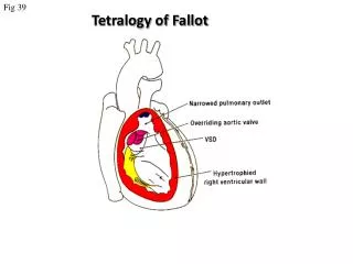
GambaranTetralogiFallot Bailliard F, Anderson RH. Tetralogy of fallot. Orphanet Journal of rare Disease. 2009; 4(2): 1-10. Webb GD, Smallhorn JF, Therrien J, Redigton AN. Congenital Disease dalamBraunwald’s Heart Disease, 9th ed. Philadephia: Elsevier Saunders; 2012. [ebook].
Embriogenesis • Park MK. Pediatric cardiology for practicioners, 4th ed. Philadelphia: Mosby; 2002. [e-book] • Apitz C, Webb GD, Redlinton AN. Tetralogy of fallot. The Lancet. 2009; 174: 1462-71.
Bailliard F, Anderson RH. Tetralogy of fallot. Orphanet Journal of rare Disease. 2009; 4(2): 1-10.
KarakteristikToF • Bailliard F, Anderson RH. Tetralogy of fallot. Orphanet Journal of rare Disease. 2009; 4(2): 1-10. • Park MK. Pediatric cardiology for practicioners, 4th ed. Philadelphia: Mosby; 2002. [e-book]
Anamnesis dan PF • Park MK. Pediatric cardiology for practicioners, 4th ed. Philadelphia: Mosby; 2002. [e-book]
Hypoxic Spell hiperpneaparoksismal (respirasidalamdancepat), iritabilitasdanprolonged crying, sianosis yang bertambah, danmenurunnyaintensitas murmur jantungkarenaabsennyaaliranantegrade yang melewatijalur RVOT. • Park MK. Pediatric cardiology for practicioners, 4th ed. Philadelphia: Mosby; 2002. [e-book]
TetralogiFallot PEMERIKSAAN PENUNJANG
PemeriksaanPenunjang • Park MK. Pediatric cardiology for practicioners, 4th ed. Philadelphia: Mosby; 2002. [e-book]
PemeriksaanLaboratorium Polisitemia Tidakterdapatdefisiensibesi
Pemeriksaan EKG ToF • Right Axis Deviation padaToFsianosis • QRS normal padapasienasianosis • Right ventricular hypertrophy • Hipertrofiventrikel bilateral • Hipertrofi atrium kanan • Park MK. Pediatric cardiology for practicioners, 4th ed. Philadelphia: Mosby; 2002. [e-book]
Hasil EKG (2 Maret 2014) Sinus rhythm, QRS rate 125x/menit, QRS normal, PR interval normal, ST-T changes (-), right axis deviation (+)
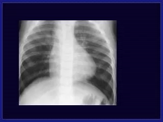
Foto X-Ray ToF • Ukuranjantung normal/lebihkecildari normal • Corakanvaskularpulmonalcenderungmenurun • Lapangparu yang “hitam” atresia pulmonal • Boot shaped appearance • Pembesaran atrium kanan & arkus aorta kanan • Park MK. Pediatric cardiology for practicioners, 4th ed. Philadelphia: Mosby; 2002. [e-book]
Hasil Rontgen (2 Maret 2014) • Jaringanlunaktidakadaemfisa • Tulang costae tidakmelebar • Tidakterdapatdeviasitrakea • Sudutkostofrenikuskanandankirilancip • Corakanbronkovaskulartidakmeningkat • CTR <50% • Tidakterdapatgambaran “boot shaped”
EkokardiografiRoF • Park MK. Pediatric cardiology for practicioners, 4th ed. Philadelphia: Mosby; 2002. [e-book]
Bailliard F, Anderson RH. Tetralogy of fallot. Orphanet Journal of rare Disease. 2009; 4(2): 1-10.
HasilEkokardiografi (14 Februari 2014) • Atrial situssolitus • AV concordance • Normal systemic and pulmonary venous drainage • Dilated RA-RV • PFO R to L shunt • TR (PG 23 mmHg) • Severe infundibulary pulmonary stenosis (PG 69 mmHg) • Large perimembranousVSD (RL shunts) • Overriding aorta ± 50% • Confluent PA’s (RPA 6 mm, LPA 3 mm) • Left aortic arch, laminar flow across • Well-contracting ventricles, no paradoxical movement seen • No pericardial effusion seen • Colateral • Kesimpulan: • Tetralogy of Fallot, smallish LPA • PFO • Colateral
Kateterisasi RFA & LFA, Plebotomi± 40 cm (12 Maret 2014) • Vena innominate (+), P-LSV C (-) • RV grafi (LAO 20, CRAN 30) • Tampakpulmonal stenosis infundibulardansupravalularberat • Overriding aorta • Confluent PA ØRPA 7,09 mm; LPA 9,01 mm • LV grafi: • Tampak VSD • Good LV Size • Aortografi: • Two ostiumcorronary artery • Left aortic arch • Tidakada PDA • Minimal kolateral • Tidakadakoartasio aorta • ØAorta desendens: 11,41 mm
Kateterisasi RFA & LFA, Plebotomi± 40 cm (12 Maret 2014) • StudiHemodinamik • Half Size: 6,5 • McGoon Ratio: 1,41 • Nakata Index: 277 • Waktuprosedur: 45 menit • Waktufuloroskopi: 3,4 menit • Kesimpulan: • Tetralogy of Fallot • Confluent PA • Normal Coronary Artery • No PDA • Minimal collateral
TetralogiFallot DIAGNOSIS
Assessment VATER • Vertebral anomalies • Anal atresia • Cardiac defects • Tracheoesophageal fistula • Renal & radial anomalies • Limb defects • Simona P, Gluffire M. Esophageal atresia in newborns: a wide spectrum from the isolated forms to a full VACTERL phenotype. Ital J Pediatr. 2013; 39(1): 1-8.