Download
1 / 10
100 likes | 397 Views
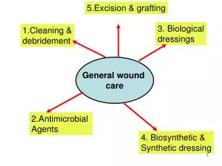
5 key Assessment areas in wound care. Disease Processes: Diseases that affect circulationarteriosclerosis venous insufficiency hypertension hyperlipidemia obesity, diabetes mellitus malignant neoplasms, All of the above can interfere with nutrition and oxygenation of cells. As they diminish t

E N D
1. Wound Care in the Emergency Dept Complete patient assessment.
This includes a health history with medical diagnosis and a physical examination to gather comprehensive, accurate data.
2. 5 key Assessment areas in wound care Disease Processes: Diseases that affect circulation
arteriosclerosis
venous insufficiency
hypertension
hyperlipidemia
obesity, diabetes mellitus
malignant neoplasms,
All of the above can interfere with nutrition and oxygenation of cells. As they diminish the body�s ability to transport leukocytes and macrophages, the immune response for controlling infection is also impaired.
3. 5 key Assessment areas in wound care Medications:
Past and current medication use, including anticoagulants, corticosteroids, immunosuppressives and antineoplastics, may adversely affect wound healing.
Nutrition and Hydration:
Malnutrition deprives the body of protein and calories required for cell growth and repair. Dehydration, by reducing blood pressure, and overhydration, which increases the distance within intracellular spaces, can impair the transport of oxygen and nutrients.
Factors that mark the possibility of malnutrition include
a recent weight loss of 10% of usual body weight
NPO status for more than three days with or without IV fluid support
problems such as malabsorption syndromes, draining wounds or fistulae, infection, or fever
4. 5 key Assessment areas in wound care Laboratory Data:
Serum Albumin (normal 3.5 to 5.0 g/dl) � Albumin, important for regenerating tissue for wound healing, comprises more than 50% of total serum protein. A low level may indicate that cells are in a destructive or catabolic state, which can lead to tissue necrosis and infection.
Serum Total Protein (normal 6.0 to 8.0 g/dl) � Low values are associated with reduced colloid osmotic pressure, so that fluids, especially plasma, are not flowing into the cells. A decline in flow leads to poor oxygenation and cell nutrition, and tissue edema.
Serum Transferrin (normal 180 to 260 mg/dl) � Transferrin is a glycoprotein that helps transport iron in the plasma, where it is required for oxygen transport to cells and for collagen synthesis. Because most iron is transported to the bone marrow for use in hemoglobin synthesis, inadequate levels may lead to anemia.
Total Lymphocyte Count (TLC) (normal 1,500 to 3,000 cells) � Some components of the immune system, such as lymphocytes, are indicators of protein status. Although a depressed TLC may indicate malnutrition, levels may also be depressed by chemotherapy, autoimmune diseases, stress, and infection.
5. Acute vs: Chronic Wound Acute
results from an injury (surgery or trauma) and progresses through the phases of wound healing in approximately one month.
a patient who is healthy and without underlying disease, healing usually occurs without complex topical treatments.1
Chronic
A chronic wound does not proceed through the phases of wound healing in an orderly or timely fashion. Underlying disease (diabetes, venous/arterial insufficiency) or external factors (pressure) contribute to the failure of the healing process.2
If a wound has not shown evidence of healing or has not healed within two weeks, it may be a chronic wound.
6. Wound Assessment Location: Describe the anatomic location of the wound to ensure accurate documentation and communication to other members of the healthcare team. Location seems to influence the rate of healing; for instance, wounds closer to the upper body usually have a greater potential for healing than wounds on the lower body.
Dimensions: Measure the length, width, and depth of the wound in centimeters for consistency in documentation. A nursing note might describe a wound as �4.5 cm L x 2 cm W x 1.5 cm D.� When measuring the depth of a wound, gently insert a sterile cotton-tipped applicator into the deepest part. Measure from the tip of the applicator to skin level. Never estimate.
Undermining and Sinus Tract Formation: Inspect ulcers, especially stage III & IV � full thickness � wounds for undermining and/or sinus tract formation. Using a sterile cotton-tipped applicator, gently probe the margins of the lesion for extensions into surrounding tissue (undermining) and beyond the wound base (for sinus tract formation). Both conditions result in dead space, open areas beneath the skin that can lead to further tissue destruction and infection.
7. Wound Assessment Tissue Viability: Healthy tissue consists of granulation tissue, which has a red, moist, beefy appearance, and epithelialized tissue - new pink, shiny epidermis. Necrotic tissue is avascular and is described as either slough or eschar tissue. Slough appears in an array of yellow, grey, green, and brown colors. Eschar is a hard, black, leathery tissue.
Exudate: Assess the exudate for volume, color, consistency, and odor. Volume of exudate is described as scant, small, moderate, or copious, and includes the number of dressings soaked with drainage. Consistent documentation allows nurses to monitor trends in wound drainage. Odor, color, and consistency of exudate can alert the nurse to the presence or absence of wound infection.
Periwound Condition: The condition of periwound skin, the area surrounding the wound opening, provides further information concerning the patient�s health status, the efficacy of a dressing�s absorption of exudate, and the presence of local infection. The nurse needs to observe this area for erythema, induration, crepitus, hematoma formation, maceration, desiccation, denudation, blistering, and pustule formation.
8. Wound Assessment Pain: Note any wound-related pain. Is the patient experiencing pain only with dressing changes or is the pain constant? How does the patient rate the pain on a scale from 1 to 5 (1 being mild, 5 excruciating)? Where is the pain? Pain may be an early symptom of infection, leading to investigation for other signs of sepsis.
Stage or Extent of Tissue Damage: For pressure ulcers, use the Wound, Ostomy and Continence Nurses (WOCN)/Agency for Health Care Policy and Research (AHCPR) criteria (See sidebar �Staging of Pressure Ulcers/Wounds�) to describe the extent of tissue damage. For other wounds, for example, vascular or diabetic, terms such as partial thickness or full thickness are useful to describe the extent of tissue damage.3
9. Staging Classification:
10. Steps to prevent Wounds in the ED Basic turn & re-position every 2 hours
Be aware of what surface your pt is lying on
Traumas on slide boards for a prolonged period
Person on longboards for prolonged periods
Admitted patients to get regular hospital beds when they will be in department for prolonged periods.
Nutritional consults for admitted pateints (Can be entered in by POE) Nurse driven initiative