Download
1 / 35
350 likes | 498 Views
Introduction. Orthopaedic Surgery is a Specialty evolved after WW I Heritage of Orthopaedic surgery is TRAUMA. Definitions. Fracture: discontinuity of cortex Union Bone restored in terms of mechanical stability Delayed Union

E N D
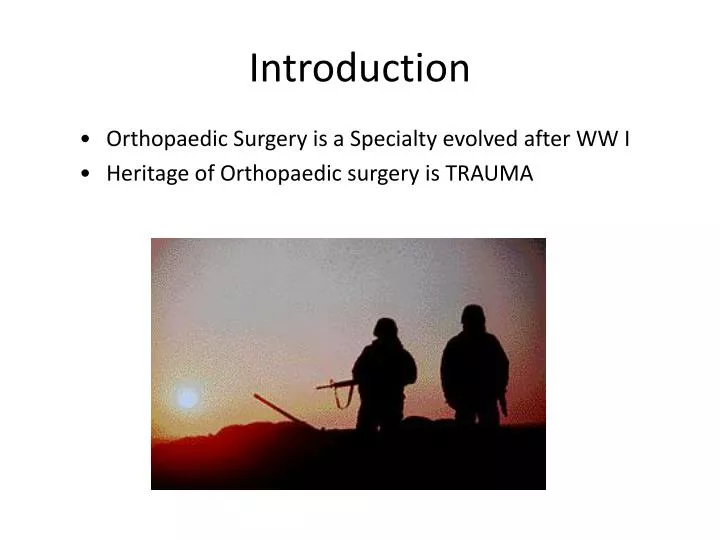
Introduction • Orthopaedic Surgery is a Specialty evolved after WW I • Heritage of Orthopaedic surgery is TRAUMA
Definitions • Fracture:discontinuity of cortex • Union • Bone restored in terms of mechanical stability • Delayed Union • Fracture is not consolidated at 3 months, but appears to be moving in that direction • Non Union • No improvement clinically or radiographically over 3 month period • Malunion
How frequent? • The overall fracture incidence is 11 in 1,000 per year. • Trauma is the Leading cause of death in < 45 age group
How does fracture occur ? • “High Energy" • Energy imparted into the bone disrupts the soft tissue envelope as a very destructive process • “Low Energy“ • Less energy imparted into the fracture environment, thus a less destructive process
How does fracture occur ? Direct trauma
How does fracture occur ? Indirect trauma
How does fracture occur ? Muscle violence
Types of fractures Direction of loading • Bending • Axial Loading • Tension • Compression • Torsion Bending Compression Torsion
Biology of Bone Healing • Primary bone healing • RequiresAbsolute Stability (rigid internal fixation) and intimate cortical contact • Relies on Haversian remodeling with bridging of small gaps by osteocytes
Biology of Bone Healing • Secondary Bone Healing = CALLUS formation (Relative Stability)
Clinical features 1) Pain
Clinical features 2) Swelling
Clinical features 3) Deformity 4) Inability to use the fractured part
Radiologic evaluation Plain X ray • 2 planes : AP & Lateral • 2 joints: above and below • 2 sides: in children • 2 occasions: scaphoid
Radiologic evaluation How to describe? Types of displacement • Angulation • Translation • Rotation • Shortening
Radiologic evaluation • CT scan
Radiologic evaluation • MRI
TREATMENT 1) First aid treatment 2) Definitive treatment
First aid treatment 1) Assess general condition: A- Airway B- Breathing C-Circulation
First aid treatment 2) Splint
Definitive treatment • Reduce: closed vs open • Hold: external vs internal • Move: rehabilitation
Closed Reduction • All displaced fractures should be reduced to minimize soft tissue complications, including those that require ORIF
Closed Reduction • Adequate analgesia and muscle relaxation are critical for success • Correct length, rotation, and angulation • Immobilize joint above and below
Closed Reduction • Reduction maneuver may be specific for fracture location and pattern • Reduction may require reversal of mechanism of injury
1) Splinting Methods of External Fixation • Non-circumferential – allows for further swelling
1) Splinting Methods of External Fixation • Types: Ulnar gutter, Volar / Dorsal hand, Thumb spica, Posterior slab (ankle), U splint
2)Casting Methods of External Fixation • More rigid immobilization • Often a poor choice in the treatment of acute fractures due to swelling and soft tissue complications
2)Casting Methods of External Fixation • Cast padding • Roll distal to proximal • 50 % overlap (2 layers minimum) • Extra padding at bony prominence
Methods of External Fixation 2)Casting • Stockinette- may require two different diameters to avoid over tight or loose material • Cast padding
Methods of External Fixation 2)Casting • Cast Molding • Mold applied to produce three point fixation • Avoid molding with anything but the heels of the palm to avoid pressure points
Methods of External Fixation 2)Casting • Complications of Casts & Splints • Tight cast post cast oedema & compartment syndrome • Loss of reduction • Pressure necrosis • Joint stiffness
3) Traction Methods of External Fixation • Allows constant controlled force for stabilization of bone fractures • It may aid in reduction during operative procedure