Download

1 / 38
410 likes | 957 Views
Iliofemoral Joint (Hip) Presented By: Angela, Clifford, Casey. Hip Joint Cut & Labeled. A. Ligaments. Iliofemoral ligament #3 Pubofemoral ligament #4. ® Anterior view (both). A. Ligaments. Ischiofemoral ligament #5. ® Posterior view. A. Ligaments.

E N D
A. Ligaments • Iliofemoral ligament #3 • Pubofemoral ligament #4 ® Anterior view (both)
A. Ligaments • Ischiofemoral ligament #5 ® Posterior view
A. Ligaments • Ligament of the head (aka – ligamentum teres femoris)
A. Ligaments • Transverse ligament of the acetabulum • Zona orbicularis
A. Ligaments • Sacrotuberous ligament • Inguinal ligament
B. Bursae The Gluteofemoral bursa is found here under the muscle • Trochanteric bursa • Ischial Bursa • Gluteofemoral bursa (aka – intermuscular bursa)
F. Clinical Concerns • Piriformis Syndrome How to get rid of a pain in the buttocks. It is thought to be caused by an impingement of the sciatic nerve by the shortening or tightening of the piriformis muscle.
G. Surface Anatomy • Gluteal fold (aka - Gluteal sulcus) • Intergluteal cleft
G. Surface Anatomy • Supracristal Plane (SCR) This is best described as a transverse plane passing through the tops or summits of the iliac crests; it usually passes through the fourth lumbar spinous process.
G. Surface Anatomy • Posterior Superior Iliac Spine
G. Surface Anatomy • Sacrum • Anterior superior iliac spine • Iliac crest
G. Surface Anatomy • Tip of Greater Trochanter • Inguinal ligament
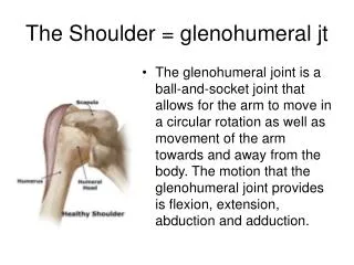
The Hip (Iliofemoral) Joint is a diarthrosis, or synovial triaxial articulation permitting movement in all three planes. Structural Classification: Ball-and-socket
Cartilage Acetabular Labrum: projecting rim located on the fibrocartilaginous layer which increases depth of joint cavity Articular Cartilage of Head of Femur: opposing cartilage surface (shown in blue) Articular Cartilage Acetabular Labrum
Articular Capsule Dense collagen fiber sleeve which surrounds the joint providing protection and stabilization. Covers approximately the proximal two thirds of the neck of the femur posteriorly. Synovial Membrane: lines the joint cavity and produces synovial fluid. Fibrous Layer: attaches proximally on the hip bone to the rim of the acetebelum and the transverse acetabular ligament
The Hip Joint is composed of the articulation between the Os Coxae and the Femur. The Os Coxae is made up of three bones: the Ilium, the Ishium and the Pubis. (The Os Coxae also articulates with the Sacrum at the Sacroiliac Joint) The Acetabulum is a concave socket shared by all three bones on the lateral surface which articulates with the Head of the Femur.
Movements of the Hip Joint Circumduction and complex motions
Four Iliac Spines can be found on the margins of the Ilieum. These spines are attachment points for muscles and ligaments and include the Anterior- Superior and Inferior spines and the Posterior-Superior and Inferior spines. The very superior point of the Ileum is called the Iliac Crest. Inferior to the Iliac Crest on the medial portion is a shallow depression called the Iliac Fossa, or Wing. Just inferior to the Posterior- Inferior Iliac Spine lies the Greater Sciatic Notch.
The Ischium forms the posterior, inferior portion of the Os Coxae. The Ischial Spine projects superior to the Lesser Sciatic Notch. The Ischial Tuberosity lies at the posterior and lateral edge of the Ischium. The Ischial Ramus connects the Ischium to the Pubis. The Inferior Ramus of the Pubis meets the Ischium and extends to the Pubic Crest. Just superior to the crest the Superior Ramus connects the Pubis to the Ilieum. Together, the Ischium and the Pubis encircle the Obturator Foramen.
The Symphysis Pubis, a pad of fibrocartilage,interconnects the medial anterior surfaces of both hip bones together. The Pubic Arch is formed by the inferior rami of the Pubis and Ishium of the two sides. A male pelvis has a pubic angle of 90 degrees or less. A female pelvis has a pubic angle of 100 degrees or more.
Femur: The longest and heaviest bone in the body The rounded Head of the Femur articulates with the acetabulum of the pelvis to form the Hip Joint. The Femur runs from the Hip Joint to articulate with the Tibia to form the Knee Joint. The Neck of the Femur joins the shaft at 125 degrees.The Greater Trochanter projects laterally. The Lesser Trochanter projects posteriorly and medially. These trochanters are where large tendons attach to the Femur.
The Linea Aspera is a prominent elevation which runs along the posterior surface and serves as an attachment site for hip muscles. The Pectineal Line is located just beneath the Lesser Trochanter. The Adductor Tubercle is a bony prominance which can be found just above the medial epicondyle.
Tibia The Tibia articulates with the Femur at the Medial and Lateral Tibial Condyles. The condyles are separated by a ridge called the Intercondylar Eminence. The Tibial Tuberosity lies near the condyles and is large and rough enough to be felt through the skin. The Anterior Margin or Crest of the Tibia runs from the Tibial Tuberosity down the anterior surface.
Sites of Bony Attachment
Psoas Major • Ant. and Lat. Surface of T12-L5 • Lesser Tochanter • Hip Flexion • L2 and L3 nerve • Iliacus • Iliac fossa • Lesser trochanter • Hip Flexion • Femoral nerve Psoas Major Iliacus
Adductor Brevis • Pubis • Pectineal line and Prox. Linea aspera • Hip Adduction • Obturator nerve • Obturator and Deep femoral artery • Adductor Magnus • Ishcium and pubis • Entire linea aspera and adductor tubercle • Hip Adduction • Obturator nerve • Obturator artery
Semimembranosus • Ischial tuberosity • Post. Surface of medial condyle of Tibia • Extend hip and flex knee • Sciatic nerve • Inf. Gluteal artery • Semitendinosus • Ischial tuberosity • Anteromedial surface of Prox. Tibia • Extend hip and flex knee • Sciatic nerve • Deep femoral
Rectus Femoris • Ant. Inf. Iliac spine • Tibial tuberosity • Hip flexion, knee extention • Femoral nerve • Lat. Circumflex femoral artery • Sartorius • Ant. Sup. Iliac spine • Prox. Medial aspect of tibia • Hip flexion, abduction, lat. Rotation • Femoral nerve • Lateral circumflex femoral artery Sartorius
Adductor Longus • Pubis • Middle one-third of the linea aspera • Hip adduction • Obturator nerve • Obturator and Deep femoral artery • Gracilis • Pubis • Ant. Med. Surface of Prox. end of tibia • Hip adduction • Obturator nerve • Obturator artery Gracilis
Biceps Femoris • Long head: Ischial tuberosity • Short head: Lat. Lip of linea aspera • Fibular head • Long head: Extend hip and flex knee • Short head: Flex knee • Long head: Sciatic nerve • Short head: Common peroneal nerve • Inf. Gluteal artery
Pectineus • Sup. Ramus of pubis • Pectineal line of femur • Hip flexion and adduction • Femoral nerve • Medial circumflex femoral artery • Tensor Fascia Latae • Ant. Sup. Iliac spine • Lateral condyle of tibia • Hip flexion and abduction • Superior gluteal nerve • Superior gluteal artery
Gluteus Maximus • Post. Sacrum and ilium • Post. Femur distal to greater tochanter • Hip extension, hyperextension, lateral rotation • Inferior gluteal nerve • Superior gluteal artery • Gluteus Medius • Lateral ilium • Greater tochanter • Hip abduction • Superior gluteal nerve • Superior gluteal artery
Gluteal minimus • Lateral ilium • Ant. Surface of the greater tochanter • Hip abduction, medial rotation • Superior gluteal nerve • Superior gluteal artery • Piriformis • Internal surface of sacrum, Sacrotuberous ligament • Sup. Border of the greater tochanter • Lateral rotation, abduction, helps hold femur in acetabulum • L5, S1-S2 nerves • Sup. And Inf. Gluteal arteries