Download
1 / 1
10 likes | 95 Views
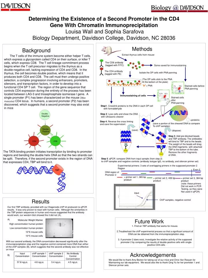
=proteins. Determining the Existence of a Second Promoter in the CD4 Gene With Chromatin Immunoprecipitation Louisa Wall and Sophia Sarafova Biology Department, Davidson College, Davidson, NC 28036. Methods. Background. Extract thymus cells from mouse.

E N D
=proteins Determining the Existence of a Second Promoter in the CD4 Gene With Chromatin Immunoprecipitation Louisa Wall and Sophia SarafovaBiology Department, Davidson College, Davidson, NC 28036 Methods Background Extract thymus cells from mouse The T cells of the immune system become either helper T cells, which express a glycoprotein called CD4 on their surface, or killer T cells, which express CD8. The T cell lineage commitment process begins when the T cell precursor migrates to the thymus as a double-negative cell, lacking expression of CD4 and CD8. In the thymus, the cell becomes double positive, which means that it produces both CD4 and CD8. The cell must then undergo positive selection, a complex progression involving enhancers, promoters, silencers, and transcription factors, in order to develop into a functional CD4 SP T cell. The region of the gene sequence that controls CD4 expression during the entirety of the process has been located between LAG-3 and triosephosphate isomerase I gene. A single promoter (P1) has been characterized on the mouse (Mus musculus) CD4 locus. In humans, a second promoter (P2) has been discovered, which suggests that a second promoter may also exist in mice. The CD8 antibody (tagged with FITC) Some saved for immunostaining The CD4 antibody (tagged with PE) Isolate the DP cells with PNA panning (The DP cells stick to the PNA At the bottom of the plate) = PNA Stained cells before PNA panning 1% 75% 6% 0.2% 81% Immunostaining of cells Stained cells after PNA panning 2% Step1. Crosslink proteins to the DNA in each DP cell with formaldehyde Double- Negative Step 2. Lyse cells and shear the DNA with Ultrasonic cleaner Step 4. Reverse the cross linking and save the supernatant. (save a portion of the sheared DNA to compare To chIP samples) Double- Positive (save) (dispose) Step 3. Add pre-blocked beads and TBP antibody. The antibodies will bind to TBP and to the beads. The weight of the beads will drag the DNA fragments with attached TBP to the bottom of the tube. Remove the other fragments of DNA. CD4 CD8 TBP TBP bead The TATA binding protein initiates transcription by binding to promoter regions and bending the double helix DNA so that the two strands can be split. Therefore, if the second promoter exists in the region of DNA that expresses CD4, TBP will bind toit. bead Step 5. qPCR: compare DNA from input sample (from step 2) to chIP samples and negative controls (antibody isotype IgG, no antibody, and silencer primer set) Experimental primers: 3 sets of overlapping primers in the supposed promoter 2 Region Exon 2 TBP DNA region of Promoter 2 = primer set 1, 365 bp product = primer set 2, 406 bp product = primer set 3, 384 bp product (note: these primers Did not work in PCR Testing, so they were Not used in qPCR) input Results ChIP samples, negative control Our first TBP antibody, provided with our Diagenode chIP kit produced no qPCR results. It was only proven to react with human cells. Although the similarities in the TBP protein sequences in human and mouse suggested that the antibody would work, our western blot showed the it did not (A). A) Future Work Molecular Weight Marker High concentration human protein 1. Find an TBP antibody that works for mouse. 2.Troubleshoot the chIP experimental process so that a significant amount of DNA can be obtained from the immunoprecipitation step. 3. If promoter 2 does exist, investigate the relative activity of the supposed promoter 2 by comparing the results of double-positive cells with single-positive CD4cells. Low concentration human protein 10^6 mouse cells 10^5 mouse cells With our second antibody, the DNA concentration decreased significantly after the immunoprecipitation step and the negative control contained more DNA than either of the chIP samples (B), which suggests that the second antibody was not effective on mice Acknowledgements B) We would like to thank Amy Becton for taking car of our mice and Chris Van Rooyen for Maintaining our lab equipment. We would also like to thank Qing Yu for her promoter 1 and Silencer primer sets.