Download

1 / 63
630 likes | 761 Views
Why cover “acuity” in a course called “Central Visual Mechanisms”? …because we are now talking about how the whole visual system works and how we can measure vision.

E N D
Why cover “acuity” in a course called “Central Visual Mechanisms”? …because we are now talking about how the whole visual system works and how we can measure vision. Acuity has a neural basis, but it is typically measured in a “whole organism” (person or animal), though it is also possible to measure the acuity of single neurons.
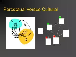
Comparison of Spatial Acuity Tasks and Thresholds Debate: are resolution and identification acuity the same?
It is an intensity discrimination task You need to be able to explain the reason that detection acuity is an intensity discrimination
All types of visual acuity are determined largely by optical and “neural” defocus For all types of acuity, need to consider these three things: The retinal image Photoreceptor sampling Convergence (receptive field center size) = “neural defocus”
How images spread out on the retina & interaction with pupil size Dashed line = theoretical point-spread function based on pupil alone (larger pupil give narrowest point-spread) Solid line = actual point-spread function based on all factors (intermediate pupil is best)
For wide objects the eye’s optics only affect the edges of the shadow’s image on the retina light At cornea dark On retina As the object gets thinner, the shadow gets thinner. BUT, when the object is smaller than 3 arc seconds (3”) the width of the shadow stays the same
The line against the sky Target Target Luminance Shadow on cornea 1.0 0.8 0.6 0.4 0.2 0.0 Retinal Illuminance 1.0 Shadows on retina 0.8 0.6 3 SEC ARC 0.4 0.2 0.0 Photoreceptor Array -1 0 +1 Retinal Position (min)
The line against the sky Target Target Luminance Shadow on cornea 1.0 0.8 0.6 0.4 0.2 0.0 Retinal 1 SEC ARC Illuminance 1.0 Shadows on retina 0.8 0.6 0.4 0.2 0.0 Photoreceptor Array -1 0 +1 Retinal Position (min)
Shadow at cornea Spread out image on retina
Shadow on cornea Shadows on retina To detect the line, the hyperpolarization of the cone at 0 must be different enough from that of adjacent cones so that the ganglion cell activity sends a strong enough signal to cortex to be detected.
In the fovea, each cone connects, through the bipolar cell, to a ganglion cell. One cone = RF center. This forms a direct line to LGN and cortex.
Shadow on cornea Shadows on retina To detect the line, the hyperpolarization of the cone at 0 must be different enough from that of adjacent cones so that the ganglion cell activity sends a strong enough signal to cortex to be detected.
Actually, a series of cones in the center of the shadow would be less hyperpolarized than the ones on either side. These cones would signal, through a bipolar cell and a ganglion cell, the presence of a line.
It is an intensity discrimination task When the shadow is so pale that the row of cones under the shadow is not hyperpolarized enough, relative to the rows of cones on each side, to cause less firing in on-center ganglion cells and more firing in off-center ganglion cells than is produce in the ganglion cells fed by adjacent rows of cones.
The retinal image Photoreceptor sampling Convergence (receptive field center size) = “neural defocus” A line needs to be thicker in the periphery to be detected because of convergence; several photoreceptors connect to a bipolar cell and several bipolar cells connect to a ganglion cell.
Vernier lines Spatial Interval
March 2007 Vision Research Vernier Acuity in the Barn Owl
The retinal image Photoreceptor sampling Convergence (receptive field center size) = “neural defocus”
The threshold for detecting the mis-alignment of lines is less than the width of a cone, so the retina cannot detect this itself. Rather, this discrimination is achieved at the cortical level (somewhere).
The Minimum Angle of Resolution (MAR) This is what is generally called “visual acuity” and is the most common measure of visual function made by eye-care practitioners
Uses of Spatial Acuity Measures • Assess if refractive error is present • Decide when to change glasses Rx • Assess visual function (best corrected refractive error) • Assess job eligibility (pilots, police, etc.) • Follow disease and treatment • Decide whether a person should drive • Decide whether a person qualifies for disability • Low vision assessment • Prediction of improvement with vision aids
Chapter 1: most of the measures of vision people make are threshold measures. All acuity measures (all three types) are threshold measures Detection acuity: we measure the threshold line width (or spot size) Localization acuity: we measure the threshold offset Resolution acuity: we measure the threshold separation
The retinal image Photoreceptor sampling Convergence (receptive field center size) = “neural defocus”
More realistic depiction of the point-spread function (line-spread function here, viewed in cross-section)
The closer together the points or lines, the less of a “dip” in intensity in between the retinal images
At the fovea, there is a match between photoreceptor size and spacing, and MAR Center-to-center spacing of 20” – 40” in the fovea Resolution 30” – 40” (0.5’) – one row of cones in between
What happens when the retinal image is defocused? Why is it that the MAR gets larger (poorer acuity) when images are out of focus? (slides from Dr. Fullard)
pupil diameter (in meters) ametropia tan (visual angle of VA chart letter) Use Blur Ratio
The closer together the points or lines, the less of a “dip” in intensity in between the retinal images
The closer together the points or lines, the less of a “dip” in intensity in between the retinal images
Convergence (receptive field center size) = “neural defocus” The larger the receptive field, the poorer the resolution acuity (lines must be spaced farther apart)