Download
1 / 160
1.63k likes | 1.95k Views
Organization of the Cell. Cell Theory. Cells are the basic living units of organization and function in all organisms and all cells come from other cells. Cell Theory. The players: Matthias Schleiden- German botanist (1838) Theodor Schwann- German zoologist (1839)

E N D
Cell Theory Cells are the basic living units of organization and function in all organisms and all cells come from other cells
Cell Theory The players: Matthias Schleiden- German botanist (1838) Theodor Schwann- German zoologist (1839) Rudolph Virchow- German professor of pathology (1855)
Schleiden and Schwann The first to point out that all plants and animals are composed of cells. 1838
Rudolph Virchow The first to observe cells dividing 1855
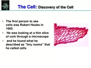
History of the Microscope • Robert Hooke examined a thin piece of cork using a compound microscope- noticed the boxes in the thin slice and called them “cells” 1665 ?
History of the Microscope Anton van Leeuwenhoek viewed living cells with 200 magnification single lenses of his own construction. His important discoveries include bacteria, protists, blood cells, and sperm cells. 1670s Dutch Scientist
2004 Nikon ‘confocal’ microscope and, “No, I don’t know how much it costs.”
Electron Microscope Invented in 1930s by (believe it or not) German scientists Max Knott and Ernst Ruska
Transmission Electron Microscope • 2-D Image • Image not living • 10,000X to 100,000X • Electron beam passes through the specimen • Specimen is thinly sliced
Scanning Electron Microscope • 3-D imaging • Image not living • 1,000X-10,000X magnification • Image is coated with a thin film of metal and the electron beams are collected as they bounce off of the specimen
Prokaryotic Cells Bacteria are prokaryotic cells. All other known organisms consist of ….. Eukaryotic Cells
Prokaryotic Cells • Structurally simpler than eukaryotic cells • Nuclear material not enclosed in a membrane • Ribosomes smaller than Euk. • Lack of membrane bound organelles
Cheek cells bacteria
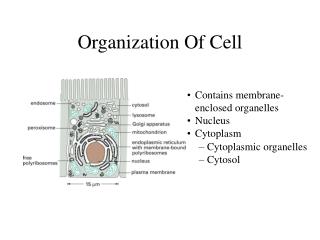
Eukaryotic Cells • Membrane bound organelles • Cell Nucleus • Ribosomes • Endoplasmic reticulum • Golgi complex • Lysosomes • Peroxisomes • Vacuoles • Mitochondria • Chloroplasts
Membrane Bound Organelles The ‘stuff’ outside the nucleus and inside the cell membrane, suspended in cytoplasm
mitochondria ribosomes Vacuoles Endoplasmic reticulum Peroxisomes Golgi complex Plastids Lysosomes Membrane Bound Organelles Just to name a few
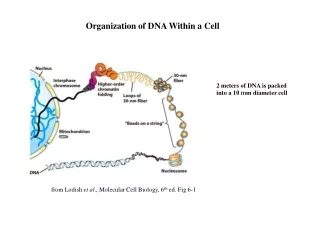
Cell Nucleus Cell Nucleus Contains nucleolus and chromosomes (DNA)
Cell Nucleus • Typically in the center of the cell • Most cells have a single nucleus
Nuclear Envelope • Controls traffic between the nucleus and the cytoplasm • Pores in the nuclear membrane allow materials to pass in and out of the • nucleus
Nuclear Lamina • Inside the nucleus • Formed by intermediate filaments • Important in the timing of the disorganization of the membrane during cell division and the ensuing redevelopment
Chromatin • When dividing, DNA takes the form of chromosomes • When not dividing, the DNA takes a looser form called chromatin
Ribosomal Subunits • Eukaryotic ribosomal subunits are assembled in the nucleolus • Ribosomes are composed of two subunits
Ribosomes • Ribosomes manufacture proteins • Ribosomes may be free or may be attached to the endoplasmic reticulum
Endoplasmic Reticulum • Major manufacturing center- proteins • Extends from the nuclear membrane into the cytoplasm • Lumen- the space enclosed by the ER- typical intracellular membrane
Rough ER • Site of protein synthesis • Proteins formed may be transferred to other sites within the cell in transport vesicles