Download
1 / 6
70 likes | 536 Views
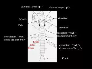
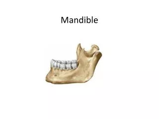
Normal Anatomic Landmarks of The Mandible. by Dorota Grela. 1. Genial tubercles- radiopaque, tiny bumps of bone that serve as attachment for genioglossus and geniohyoid muscles.

E N D
Normal Anatomic Landmarks of The Mandible by Dorota Grela
1. Genial tubercles- radiopaque, tiny bumps of bone that serve as attachment for genioglossus and geniohyoid muscles. 2. Lingual foramen-radiolucent, hole or opening located on the internal surface of mandible and surrounded by the genial tubercles. The dot appears inferior to the apices of the mandibular incisors.
1. Mandibular canal- (black arrows), tube like passage extending from mandibular foramen to the mental foramen and contains Inferior Alveolar nerv and blood vessels. This radiolucent band is outlined by two radiopaque lines of cortical plate. 2. Mental foramen- ( white circle), small ovoid radiolucent area located below the apices of the premolars. Mental foramen is located on the external surface of the mandible as an opening in the region of the mandibular premolars. Mental nerves and blood vessels exit through it.
External oblique ridge – (red arrows) radiopaque band extending downwards and forwards from anterior border of mandible and ends around third or second molar region. 2. Internal oblique ridge (Mylohyoid ridge)- ( blue arrows), radiopaque band extending downward from molars and toward the lower border of mandibular symphysis.
Submandibular gland fossa ( Submaxillary gland fossa)- nb. 13. radiolucent,depressed area located on the internal surface of mandible,in area in the molar region below the mylohyoid ridge. Submandibular salivary gland lies in this fossa.