Download
1 / 1
10 likes | 123 Views
Brain Tumor Segmentation: Label each voxel in MR image as { tumor, non-tumor } Use only individual voxels Discriminative classifier (Logistic Regression; SVMs) Also use spatial correlations of labels among neighboring voxels

E N D
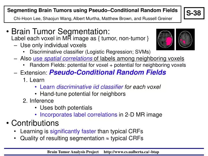
Brain Tumor Segmentation:Label each voxel in MR image as { tumor, non-tumor } Use only individual voxels Discriminative classifier (Logistic Regression; SVMs) Also use spatial correlations of labels among neighboring voxels Random Fields: potential for voxel + potential for neighboring voxels Extension: Pseudo-Conditional Random Fields Learn Learn discriminative iid classifier for each voxel Hand-tune potential for neighbors Inference Uses both potentials Incorporates label correlations in 2-D MR image Contributions Learning is significantly faster than typical CRFs Quality of resulting segmentation typical CRFs Segmenting Brain Tumors using Pseudo–Conditional Random Fields Chi-Hoon Lee, Shaojun Wang, Albert Murtha, Matthew Brown, and Russell Greiner S-38 Brain Tumor Analysis Projecthttp://www.cs.ualberta.ca/~btap