Download
1 / 128
1.28k likes | 1.29k Views
19 Blood. An Introduction to Blood and the Cardiovascular System. Learning Outcomes 19-1 Describe the components and major functions of blood, identify blood collection sites, and list the physical characteristics of blood.

E N D
19 Blood
An Introduction to Blood and the Cardiovascular System • Learning Outcomes • 19-1 Describe the components and major functions of blood, identify blood collection sites, and list the physical characteristics of blood. • 19-2 Specify the composition and functions of plasma. • 19-3 List the characteristics and functions of red blood cells, describe the structure and functions of hemoglobin, describe how red blood cell components are recycled, and explain erythropoiesis.
An Introduction to Blood and the Cardiovascular System • Learning Outcomes • 19-4 Explain the importance of blood typing, and the basis for ABO and Rh incompatibilities. • 19-5 Categorize white blood cell types based on their structures and functions, and discuss the factors that regulate the production of each type. • 19-6 Describe the structure, function, and production of platelets. • 19-7 Discuss the mechanisms that control blood loss after an injury, and describe the reaction sequences responsible for blood clotting.
An Introduction to Blood and the Cardiovascular System • The Cardiovascular System consists of: • A pump (the heart) • A conducting system (blood vessels) • A fluid medium (blood) • Is specialized fluid of connective tissue • Contains cells suspended in a fluid matrix
An Introduction to Blood and the Cardiovascular System • Blood • Transports materials to and from cells • Oxygen and carbon dioxide • Nutrients • Hormones • Immune system components • Waste products
19-1 Physical Characteristics of Blood • Important Functions of Blood • Transportation of dissolved substances • Regulation of pH and ions • Restriction of fluid losses at injury sites • Defense against toxins and pathogens • Stabilization of body temperature
19-1 Physical Characteristics of Blood • Whole Blood • Plasma • Fluid consisting of: • Water • Dissolved plasma proteins • Other solutes • Formed elements • All cells and solids
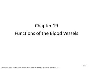
Figure 19-1 The Composition of Whole Blood 7% Plasma Plasma Proteins 1% Other Solutes 46–63% 92% Water Formed Elements Platelets < .1% White Blood Cells 37–54% Red Blood Cells < .1% 99.9%
19-1 Physical Characteristics of Blood • Three Types of Formed Elements • Red blood cells(RBCs) or erythrocytes • Transport oxygen • White blood cells(WBCs) or leukocytes • Part of the immune system • Platelets • Cell fragments involved in clotting
19-1 Physical Characteristics of Blood • Hemopoiesis • Process of producing formed elements • By myeloid and lymphoid stem cells • Fractionation • Process of separating whole blood for clinical analysis • Into plasma and formed elements
19-1 Physical Characteristics of Blood • Three General Characteristics of Blood • 38C (100.4F) is normal temperature • High viscosity • Slightly alkaline pH (7.35–7.45)
19-1 Physical Characteristics of Blood • Characteristics of Blood • Blood volume (liters) = 7% of body weight (kilograms) • Adult male 5 to 6 liters • Adult female 4 to 5 liters
19-2 Plasma • The Composition of Plasma • Makes up 50–60% of blood volume • More than 90% of plasma is water • Extracellular fluids • Interstitial fluid (IF) and plasma • Materials plasma and IF exchange across capillary walls • Water • Ions • Small solutes
19-2 Plasma • Plasma Proteins • Albumins (60%) • Globulins (35%) • Fibrinogen (4%)
19-2 Plasma • Albumins (60%) • Transport substances such as fatty acids, thyroid hormones, and steroid hormones • Globulins (35%) • Antibodies, also called immunoglobulins • Transport globulins (small molecules): hormone-binding proteins, metalloproteins, apolipoproteins (lipoproteins), and steroid-binding proteins • Fibrinogen (4%) • Molecules that form clots and produce long, insoluble strands of fibrin
19-2 Plasma • Serum • Liquid part of a blood sample • In which dissolved fibrinogen has converted to solid fibrin
19-2 Plasma • Other Plasma Proteins • 1% of plasma • Changing quantities of specialized plasma proteins • Peptide hormones normally present in circulating blood • Insulin, prolactin (PRL), and the glycoproteins thyroid-stimulating hormone (TSH), follicle-stimulating hormone (FSH), and luteinizing hormone (LH)
19-2 Plasma • Origins of Plasma Proteins • More than 90% made in liver • Antibodies made by plasma cells • Peptide hormones made by endocrine organs
19-3 Red Blood Cells • Red blood cells (RBCs) • Make up 99.9% of blood’s formed elements • Hemoglobin • The red pigment that gives whole blood its color • Binds and transports oxygen and carbon dioxide
19-3 Red Blood Cells • Abundance of RBCs • Red blood cell count - the number of RBCs in 1 microliter of whole blood • Male: 4.5–6.3 million • Female: 4.2–5.5 million
19-3 Red Blood Cells • Abundance of RBCs • Hematocrit -(packed cell volume, PCV) percentage of RBCs in centrifuged whole blood • Male: 40–54 • Female: 37–47
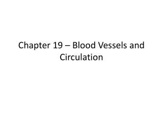
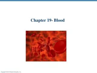
19-3 Red Blood Cells • Structure of RBCs • Small and highly specialized discs • Thin in middle and thicker at edge
19-3 Red Blood Cells • Three Important Effects of RBC Shape on Function • High surface-to-volume ratio • Quickly absorbs and releases oxygen • Discs form stacks called rouleaux • Smooth the flow through narrow blood vessels • Discs bend and flex entering small capillaries • 7.8-µm RBC passes through 4-µm capillary
Figure 19-2a The Anatomy of Red Blood Cells Blood smear LM 477 When viewed in a standard blood smear, RBCs appear as two-dimensional objects, because they are flattened against the surface of the slide.
Figure 19-2b The Anatomy of Red Blood Cells Red blood cells SEM 1838 The three-dimensional shape of RBCs
Figure 19-2c The Anatomy of Red Blood Cells 2.31–2.85 μm 0.45–1.16 μm 7.2–8.4 μm A sectional view of a mature RBC, showing the normal ranges for its dimensions
Figure 19-2d The Anatomy of Red Blood Cells Red blood cell (RBC) Rouleau (stacked RBCs) Nucleus of endothelial cell Blood vessels (viewed in longitudinal section) LM 1430 Sectioned capillaries When traveling through relatively narrow capillaries, RBCs may stack like dinner plates.
19-3 Red Blood Cells • Life Span of RBCs • Lack nuclei, mitochondria, and ribosomes • Means no repair and anaerobic metabolism • Live about 120 days
19-3 Red Blood Cells • Hemoglobin(Hb) • Protein molecule that transports respiratory gases • Normal hemoglobin (adult male) • 14–18 g/dL whole blood • Normal hemoglobin (adult female) • 12–16 g/dL whole blood
19-3 Red Blood Cells • Hemoglobin Structure • Complex quaternary structure • Four globular protein subunits • Each with one molecule of heme • Each heme contains one iron ion
19-3 Red Blood Cells • Hemoglobin Structure • Iron ions • Associate easily with oxygen (oxyhemoglobin, HbO2) • Dissociate easily from oxygen (deoxyhemoglobin)
Figure 19-3 The Structure of Hemoglobin chain 1 chain 1 chain 2 Heme Heme chain 2 Hemoglobin molecule
19-3 Red Blood Cells • Fetal Hemoglobin • Strong form of hemoglobin found in embryos • Takes oxygen from mother’s hemoglobin
19-3 Red Blood Cells • Hemoglobin Function • Carries oxygen • With low oxygen (peripheral capillaries) • Hemoglobin releases oxygen • Binds carbon dioxide and carries it to lungs • Forms carbaminohemoglobin
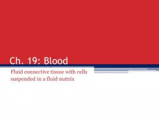
19-3 Red Blood Cells • RBC Formation and Turnover • 1% of circulating RBCs wear out per day • About 3 million RBCs per second • Hemoglobin Conversion and Recycling • Macrophages of liver, spleen, and bone marrow • Monitor RBCs • Engulf RBCs before membranes rupture (hemolyze)
19-3 Red Blood Cells • Hemoglobin Conversion and Recycling • Phagocytes break hemoglobin into components • Globular proteins to amino acids • Heme to biliverdin • Iron
19-3 Red Blood Cells • Hemoglobin Conversion and Recycling • Hemoglobinuria • Hemoglobin breakdown products in urine due to excess hemolysis in bloodstream • Hematuria • Whole red blood cells in urine due to kidney or tissue damage
19-3 Red Blood Cells • Breakdown of Biliverdin • Biliverdin (green) is converted to bilirubin (yellow) • Bilirubin • Is excreted by liver (bile) • Jaundice is caused by bilirubin buildup • Converted by intestinal bacteria to urobilins and stercobilins
19-3 Red Blood Cells • Iron Recycling • Iron removed from heme leaving biliverdin • To transport proteins (transferrin) • To storage proteins (ferritin and hemosiderin)
Figure 19-5 Recycling of Red Blood Cell Components Events Occurring in the Red Bone Marrow Events Occurring in Macrophages RBC formation Macrophages in liver, spleen, and bone marrow Fe2+ transported in circulation by transferrin Fe2+ Amino acids Heme Average life span of RBC is 120 days 90% Biliverdin New RBCs released into circulation Old and damaged RBCs Bilirubin 10% In the bloodstream, the rupture of RBCs is called hemolysis. Bilirubin bound to albumin in bloodstream Hemoglobin that is not phagocytized breaks down, and the alpha and beta chains are eliminated in urine. Liver Kidney Bilirubin Hb Absorbed into the circulation Urobilins Excreted in bile Urobilins, stercobilins Eliminated in urine Bilirubin Events Occurring in the Kidney Eliminated in feces Events Occurring in the Liver Events Occurring in the Large Intestine
Figure 19-5 Recycling of Red Blood Cell Components Events Occurring in Macrophages Macrophages in liver, spleen, and bone marrow Fe2+ transported in circulation by transferrin Fe2+ Amino acids Heme Average life span of RBC is 120 days 90% Biliverdin Old and damaged RBCs Bilirubin 10% In the bloodstream, the rupture of RBCs is called hemolysis. Bilirubin bound to albumin in bloodstream Hemoglobin that is not phagocytized breaks down, and the alpha and beta chains are eliminated in urine.
Figure 19-5 Recycling of Red Blood Cell Components Events Occurring in the Red Bone Marrow RBC formation Fe2+ transported in circulation by transferrin Average life span of RBC is 120 days New RBCs released into circulation In the bloodstream, the rupture of RBCs is called hemolysis. Hemoglobin that is not phagocytized breaks down, and the alpha and beta chains are eliminated in urine.
Figure 19-5 Recycling of Red Blood Cell Components Events Occurring in the Liver Bilirubin bound to albumin in bloodstream Hemoglobin that is not phagocytized breaks down, and the alpha and beta chains are eliminated in urine. Liver Bilirubin Absorbed into the circulation Excreted in bile Urobilins, stercobilins Bilirubin
Figure 19-5 Recycling of Red Blood Cell Components Events Occurring in the Kidney Hemoglobin that is not phagocytized breaks down, and the alpha and beta chains are eliminated in urine. Kidney Hb Absorbed into the circulation Urobilins Events Occurring in the Large Intestine Urobilins, stercobilins Eliminated in urine Bilirubin Eliminated in feces
19-3 Red Blood Cells • RBC Production • Erythropoiesis • Occurs only in myeloid tissue (red bone marrow) in adults • Stem cells mature to become RBCs
19-3 Red Blood Cells • Hemocytoblasts • Stem cells in myeloid tissue divide to produce: • Myeloid stem cells become RBCs, some WBCs • Lymphoid stem cells become lymphocytes
19-3 Red Blood Cells • Stages of RBC Maturation • Myeloid stem cell • Proerythroblast • Erythroblasts • Reticulocyte • Mature RBC
Figure 19-6 Stages of RBC Maturation RED BONE MARROW Day 1: Proerythroblast Erythroblasts Day 2: Basophilic erythroblast Day 3: Polychromatophilic erythroblast Day 4: Normoblast Ejection of nucleus Days 5–7: Reticulocyte Enters circulation Mature red blood cell
19-3 Red Blood Cells • Regulation of Erythropoiesis • Building red blood cells requires: • Amino acids • Iron • Vitamins B12, B6, and folic acid • Pernicious anemia • Low RBC production • Due to unavailability of vitamin B12