Download
1 / 20
200 likes | 320 Views
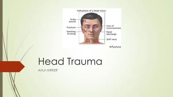
Head Trauma. Dr. Roberts. Epidemiology. 1.1 million annual ED visits Highest < 5 yo & >85 yo 80% minor head trauma (GCS 14-15) 10% moderate (GCS 9-13) & 10% severe (8 & below) 200,000 deaths, most under 25 yo & 40% firearm related & 34% MVC. Anatomy.

E N D
Head Trauma Dr. Roberts
Epidemiology • 1.1 million annual ED visits • Highest < 5 yo & >85 yo • 80% minor head trauma (GCS 14-15) • 10% moderate (GCS 9-13) & 10% severe (8 & below) • 200,000 deaths, most under 25 yo & 40% firearm related & 34% MVC
Anatomy • Brain covered in multiple layers: 1. dura 2. arahnoid 3. pia • Subarchnoid space contains 150cc CSF; 500 cc made each day • Normal CSF pressures 5-15 mmHg • Scalp 1. skin 2. subcutaneous, 3. galea, 4. areolar 5. pericranium • rich blood supply
Pathopphysiology • Two main mechanisms of injury • Primary: initial mechanical trauma (irreversible) • Secondary: hypotension; hypoxia; anemia (our job) • Cushings Reflex:Hypertension; bradycardia; respiratory irregularity • Cerebral herniation: • Central Transtentorial-expanding lesion at frontal or occipital poles; AMS, pinpoint pupils, bi-muscle weakness • Cerebellotonsillar-cerebellar tonsils herniate through foramen magnum due to cerebellar mass; Pinpoint pupils, quadriplegia and cardiorespiratory collapse • Upward Transtentorial-expanding posterior fossa lesion; pinpoint pupils, absence of vertical eye movements • Uncal-most common, usually due to hematoma, 3rd nerve compression (anisocoria, ptosis, sluggish pupil, CN III defects)
Types of herniation • Upward Transtentorial • Central Transtentorial • Uncal • Cerebellotonsillar
Initial ED Evaluation & Tx • History: • High Risk – prolonged amnesia, anticoagulation, coagulopathy, progressive vomiting, post injury seizure • Physical Exam-Neuro Exam (GCS) • High Risk – focal neuro findings, distracting injury, signs of skull fracture, large extracranial hematoma, intoxication • ABCs (consider lidocaine if RSI) • Maintain PO2 & MAP • Watch for cushings • CT if GCS < 14, high risk Hx or Exam
Further ED Management • Indications for Seizure Prophylaxis • Depressed skull fracture • Paralyzed & Intubated patient • Seizure at time of injury • Seizure in ED • Penetrating brain injury • GCS <9 • Acute Subdural/Epidural hematoma • Intracranial hemorrhage • Prior history of seizure
Specific Head Injuries • Scalp Lac: direct pressure, lido with epi, explore wound, suture/staples
Skull Fractures • Linear & simple comminuted fx: irrigate, suture, antibiotics per neuro surg consult • Basilar: CSF otorrhea/rhinorrhea, battles, raccoon, hemotympanum, vertigo, CN VII palsy, deafness, antibiotics usually not warrented
Specific Injuries • Cerebral Contusion: frequent injury, coup vs contre-coup, often with subarachnoid bleed, initial scans may be normal
Subarachnoid Hemorrhage • Disruption of small subarachnoid vessels • Only detected 33% on initial CT • Most common abnormality on Head CT • Show signs of photophobia & headache • Marks significant increase morbidity/mortality in severe head injury
Subdural Hematoma • Blood clot between dura and brain • Seen in acceleration-deceleration injuries • Common in alcoholic & elderly • Rupture of superficial bridging vessels • Acute-symptoms in 1st 24 hrs (lucid interval) • Subacute-symptoms between 24 hrs-2 wks • Chronic-symptoms after 2 wks
Epidural Hematoma • Collection of blood between skull & dura due to blunt trauma causing rupture of middle meningeal artery • May have a lucent period following immediate LOC • Due to arterial bleeding, herniation occurs quickly
Concussion • Temporary & brief interruption of neurologic function after minor trauma • Symptoms-headache, confusion, & amnesia • Should not return to play until resolution of symptoms for 1 week
Pediatric Head Trauma • <2yo consider abuse • Higher mortality in children • <3months asymptomatic, no scalp hematoma then no CT • 3months-2yrs: scalp hematoma present then skull films, if fracture CT • >2yrs CT if high risk PE or history
Penetrating Head Injuries • ABCs • Antibiotics & Td proph • CT & Neurosurgery