Download
1 / 56
560 likes | 566 Views
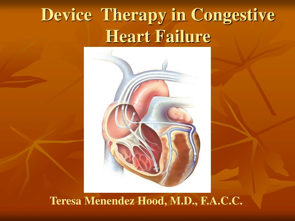
Device Therapy in Congestive Heart Failure. Teresa Menendez Hood, M.D., F.A.C.C. Congestive Heart Failure. Up to 30 % of CHF patients have an IVCD (80% with a LBBB) which has been linked to increases in mortality and morbidity.

E N D
Device Therapy in Congestive Heart Failure Teresa Menendez Hood, M.D., F.A.C.C.
Congestive Heart Failure • Up to 30 % of CHF patients have an IVCD (80% with a LBBB) which has been linked to increases in mortality and morbidity. • CHF is the leading cause hospitalizations in the US and uses up 5% of the health care costs (1999 stats) • 1-2% of the population and 6% of the population >65 • Prevalence is on the rise. Annual Incidence Heart Failure Prevalence Annual Mortality 5.0 million 400,000 250,000 U.S.
NYHA Class-evaluates the disability imposed on the patient • The FDA and the ACC/AHA Guidelines have approved biventricular pacing for class 3 and 4. Class I Asymptomatic heart failureejection fraction (EF) <40% Class II Mild symptomatic heart failure with ordinary exertion Class III Moderate symptomatic heart failure with less than ordinary exertion Class IV Symptomatic heart failure at rest
Leading Causes of Death in the U.S. Septicemia You must combine deaths from all cancers to outnumber the deaths from SCA each year. Nephritis Alzheimer’s Disease Influenza/pneumonia Diabetes Accidents/injuries Chronic lower respiratory diseases Cerebrovascular disease Other cardiac causes Sudden cardiac arrest (SCA) All other causes All cancers 0% 5% 10% 15% 20% 25% National Vital Statistics Report. Oct. 12, 2001;49(11). MMWR. State-specific mortality from sudden cardiac death – US 1999. Feb 15, 2002;51:123-126.
SCD Rates in CHF Patients with LV Dysfunction 45 months 13 months 41.4 months 27 months 12 months 16 months 6 months Total Mortality ~15-40%; SCD accounts for ~50% of the total deaths.
SCD in Heart Failure • QRS duration is an independent predictor of mortality (>140 ms) • Other factors are: age, creatinine, EF, and HR QRS 100% Duration (msec) <90 90% 90 - - 120 Cumulative Survival 80% 120 - - 170 170 - - 220 70% >220 60% 0 60 120 180 240 300 360 Days .
SCD in Heart Failure • Degree of SCD risk by class • Mortality in NYHA class II is 5 to 15% • 50 to 80% of the deaths are Sudden • Mortality in NYHA class III is 20 to 50% • Up to 50% of the deaths are Sudden • Mortality in NYHA class IV is 30 to 70% • 5 to 30% of deaths are Sudden
Right Ventricular Pacing • RV apex pacing is harmful in patients with LV dysfunction • “Paced” LBBB • Abnormal LV activation • Reduced stroke volume
RV pacing • MADIT II (2002) had a survival benefit with the ICD but in a subgroup analysis, there was an increase in heart failure morbidity (more hospitalizations) felt due to forced RV pacing compared to controls in which no pacing was present.
(p= 0.09) N= 490 N= 742 MADIT II: ComplicationsNew or Worsening HF • RV pacing causes ventricular dysynchrony and may lead to worsening HF. • Intrinsic ventricular activation is better for ICD patients with left ventricular dysfunction who do not “need” pacing. • <10% of ICD patients have a Class I pacing indication at the time of implant. • Physicians, when appropriate, should consider programming of ICDs to avoid frequent RV pacing.
DAVID — Dual Chamber and VVI Implantable Defibrillator Trial • ICD indication but no indication for a pacemaker • Ef < 40% • DDDR @ 70BPM versus VVI 40 BPM
TheConcept • In most patients with an IVCD (QRS > 130 ms) , the presence of atrial-biventricular (RV + LV) pacing will provide early stimulation to an otherwise late segment of electrical activation in the LV. • This should translate into an increase in the EF, decrease of the LV dimension, improvement in the QOL and NYHA class. • This may translate into an decrease in CHF exacerbations , hospitalizations and a decrease in mortality.
The Proof • 1994–1997: Mechanistic and both short- and longer-term observational studies. Studies initially used epicardial leads placed by thoracotomy or thorascope. • The first BiV pacer was implanted in 1994 • 1998–1999: Randomized, controlled studies to assess exercise capacity, functional status, and quality of life. • There was development of transvenous leads via the coronary sinus in to get to the LV. Cohen TJ, Klein J. J Inva2002;14:48-53.
The Proof • 2000–2005: Randomized, controlled trials to assess combined mortality and CHF hospitalization. Also evaluated the combined benefit of ICD’s with CRT. • Future: Identify patients who will benefit from CRT along with the QRS duration.This will use echocardiographic markers of asynchrony. • 20% of patients do not respond to therapy in clinical trials with a wide QRS and 50% patients with a narrow QRS/CHF have asynchrony on echo and may benefit from this therapy. • If the QRS is < 150 then the chances of responding to BiVP is ~5%. It will be in this patient group of QRS of 120-150 ms where preselection of responders will be most valuable.
The Cardiac Resynchronization Clinical Trials PATH-CHF, MUSTIC, MIRACLE, COMPANION, and CARE-HF* *This is not a complete list of all the CRT trials and the dates given are when the trial results were published.
Cumulative Enrollment in Cardiac Resynchronization Randomized Trials
PATH-CHF: 1999 Pacing Therapy for Congestive Heart Failure • This was the first multicenter trial and used the standard endocardial RV lead and an epicardial LV lead via thoracotomy or thorascope • Single blinded RCT • 53 centers in Europe • 41 patients
NYHA class III-IV DCM QRS > 120 ms PR>150 Implant Acute hemodynamic testing Randomization 1:1 Best single chamber CRT 4 weeks No CRT No CRT 8 weeks CRT Best single chamber 12 weeks Best mode One year PATH-CHF
PATH-CHF • Primary endpoints • Peak VO2 • Six-minute walk distance • Secondary endpoints • Minnesota Living with Heart Failure score (QOL) • NYHA class • EF • Trend towards decrease in Hospitalizations • Acute hemodynamic testing revealed that the lateral and posterolateral walls were the best target sites. • The best responders were those with QRS>150 , long PR and dP/dt < 700 mm Hg/s
MUSTIC: 2001Multicenter Stimulation in CM • European study with 67 patients • QRS>150, CHF, EF <35% • BiVP versus backup VVI pacing at 40 BPM • Increase in 6 minute walk time , QOL and Peak VO2 with BiVP and persisted for up to 12 months • 60% decrease in CHF hospitalizations • First to use endocardial LV leads via the CS • No significant change in mortality, but a trend towards an improvement. • Acute hemodynamic studies showed the mid lateral wall to be the best site
MIRACLE:2002Multi-center In Sync Randomized Clinical Evaluation Trial • Double blinded RCT • First US trial • Class 3 or 4, on OPT, QRS >130 ms, EF<35% • Enrollment of 453 patients
NYHA class III-IV LVEDD > 60 mm QRS > 130 ms Stable 3 month regimen of beta-blocker/ACE inhibitor EF < 35% Randomization CRT on CRT off 1- and 3-month follow-up 1- and 3-month follow-up 6-month follow-up 6-month follow-up CRT on Long-term follow-up MIRACLE
P < 0.001 67% 60% 40% Proportion 39% 34% 27% 20% 17% 16% 0% Improved No Change Worsened Control N=225 CRT N=228 MIRACLE Nonresponders: older, ischemic CM, no MR, QRS<150 Responders: had shorter duration on CHF and longer QRS>155
MIRACLE • There was a decrease in hospitalizations of 50% at 6 months and a trend towards a decrease in mortality. • All other primary and secondary endpoints were met: 6 minute walk time, peak Vo2, QOL, EF , NYHA class, LVEDD Magnitude of improvement not influenced by degree of QRS shortening with BiVP (average in all was –20msec)
FDA Approval • The first CRT device was approved by the FDA in September 2001 . • The first CRT with an ICD was approved by the FDA in May 2002 .
The primary ICD prevention trials • MADIT 1 1996required a positive EP study • MUSTT1999 required a positive EP study • Madit 22002 prior MI (ischemic cardiomyopathy) and EF<30% (no EP study required) ;60% had CHF and 50% had QRS > 120 ms; resulted in a 31% decrease risk of death and halted prematurely due to the positive effect of the ICD: resulted in the FDA approving the ICD for primary prevention this patient population, but only those with a QRS > 120 ms.
The primary ICD prevention trial • SCD-Heft 2005 The SCD-Heft trial resulted in FDA approval of the ICD January 2005 in patients with CHF and EF<35 % that included both ischemic and nonischemic cardiomyopathy for primary prevention without a positive EP study or ventricular ectopy . No QRS cutoff was required.
ACC/AHA/NASPE 2002 Indications for Cardiac Resynchronization Therapy • Class II a ( Level A) Indication for Biventricular Pacing in Dilated Cardiomyopathy • Biventricular pacing in medically refractory, symptomatic NYHA Class III/IV patients with idiopathic dilated or ischemic cardiomyopathy, prolonged QRS interval (130 msec), LV end diastolic diameter 55mm, and LVEF 35%
1 • OPT 2 + • OPT • CRT Randomization 2 • OPT • CRT-D + COMPANION:2004 Comparison of Medical Therapy, Pacing and Defibrillation in Heart Failure
COMPANION • Enrolled 1520 patients class 3 and 4, QRS >120ms • Primary endpoint: death or hospitalization for any cause • CRT met the primary endpoints and the CRT +/- ICD significantly reduces mortality • This was the first to show mortality benefit from CRT alone • Showed that patients with CRT also benefit from ICD therapy • OPT had SCD in 36%,23% in CRT and 2.9% in CRT+ICD
COMPANION • CRT arm had 20% reduction in mortality and hospitalization over OPT arm but it was not statistically significant • Significant reduction in CRT-ICD arm of 40% for mortality over OPT arm (19% in OPT and 11% in CRT-ICD group) • Study was halted prematurely due to its positive benefit. • Mean follow up was 16 months
CARE-HF : March 2005 • The effect of cardiac resynchronization on morbidity and mortality in heart failure in 813 patients in Europe ( prospective multicenter RCT) with completed enrollment by 2002 • Large patient size and length of trial (average follow up of 29 months) allowed ability to asses effects of CRT • Looked at CRT alone (no ICD) • Patients with class 3 or 4, EF < 35%, QRS >120 ms • There was a 37% reduced mortality or first hospitalization for a cardiac cause compared to OPT
CARE-HF • All endpoints were met : EF, NYHA, QOL, BNP, Echo and hemodynamic parameters • 33% of the deaths in the CRT group were due to SCD • For every 9 devices, one death and 3 hospitalizations were prevented • Echo criteria in patients with QRS 120-149ms to look for asynchrony (had to have 2 of 3) • Aortic pre-ejection delay of > 140 ms ( onset of QRS to Aortic ejection) • Interventricular mechanical delay of >40 ms ( RV-LV) • Delayed activation of the postero-lateral LV wall (>50ms)
Anatomical Challenges • Enlarged right atrium • Abnormal CS location • Presence of valves in CS • Altered CS angulation • Acute branch take offs • Tortuous vessel anatomy
CRT Procedure and Device Related Risks relative to CS placement • CS lead dislogdement 8% • CS dissection or perforation 5% • Failure of lead placement 8% • Phrenic nerve stimulation 2% • ALL other risks associated with pacer or ICD implantation and anesthesia in these patients.
CS Leads they now come in many shapes and sizes and the the OTW system
Right AtrialLead Left VentricularLead Right VentricularLead Achieving Cardiac Resynchronization Goal: Atrial synchronous biventricular pacing Transvenous approach for left ventricular lead via coronary sinus Back-up epicardial approach
BASE Anterior Posterior APEX RAO is best to distinguish BASE position from APEX
ANTERIOR Anterior LAO is best to distinguish LATERAL position from SEPTAL LATERAL SEPTAL Posterior Lateral INFERIOR
The implant • 3 separate sticks via Seldinger technique in the subclavian vein -can be done from the right but it is more difficult. • Use standard peel back sheaths for the RA and RV leads • The RV lead is positioned first - could develop CHB or VT so it is good to have this in (screw-in or tined) • Advance the long guide sheath into the RA ( not to the CS) • Advance a Coronary Sinus EP catheter via the long guide sheath into the CS – the LAO is the best: point towards the spine. • Advance the sheath while pulling back on the CS catheter to get the sheath into the CS • Some would use dye at this point to look at the anatomy of the CS and its branches
The implant • Advance the CS lead with or without the OTW system and make sure you place it in a mid/lateral or posterolateral position. Never go where the LAD would be but where the obtuse marginals would be. • Test the CS lead including at 10 volts for phrenic nerve stimulation • Pull back on the sheath until it is out of the OS, then peel it out with a retention guide wire in the CS-be careful about dislodgement • Position the atrial lead in the RAA (screw-in or tined) • Test the ICD with induction of VF twice separated by 3-5 minutes: can do at a later time if the time is > 4 hours or the patient has been unstable in any way. Always use a high energy device in these patients.
The 3 levels of asynchrony • Intraventricular asynchrony is best treated by placing the LV lead in the best anatomiclocation-usually the lateral or posterolateral (proven my multiple studies). Get the LV working. • Interventricular asynchrony is dealt with by adjusting the V-V interval. Get the RV and the LV to work together. • A-V asynchrony is dealt with by adjusting the A-V interval. Get the atria and the ventricles working together.
Posterolateral or Lateral walls are the best with LBBB where the septum contracts first and then the lateral wall last. Paced at most mechanically delayed LV site Paced at any other LV site 0 10% P=0.04 -5 -9.2 8% 9% -10 6% Improvement -15 4% -20 2% -25 -28.4 P=0.04 2% -30 0% Change in LV End-systolic Volume [ml] Change in LVEF [%]
CRT and Tissue Doppler Imaging -a measure of intraventricular delay • Measures dyssynchronous (delayed) contraction patterns @ different areas of the ventricle • Measure from the onset of the QRS to the peak systolic shortening of that segment • Defined as a segment with > 50 ms delay: this indicates intraventricular delay or asynchrony by ECHO criteria • Colors: green-yellow-red (the longest delay of >300 ms)
AV Delay Optimization Methods • Electrocardiographic • COMPANION trial method • Echocardiographic (combined) • Aortic velocity time integral (VTI) methods • Mitral velocity Doppler methods:E and A waves • Ritter formula • Hemodynamic measurements • Pulse pressure method • dP/dtmax method
COMPANION Method: QRS < 150 Intrinsic QRS duration: QRS = 140 ms Intracardiac AV interval: AS to VS = 300 ms Sensed AV Delay:
COMPANION Method: QRS >150 Intrinsic QRS duration: QRS = 180 ms Intracardiac AV interval: AS to VS = 240 ms Sensed AV Delay:
Aortic VTI Method • Objective: • Identify the AV Delay that yields the maximum cardiac output as determined by an aortic VTI measurement • Procedure: • Obtain continuous wave Doppler echo of aortic valve outflow to obtain VTI measurement • Record VTI values over a range of programmed AV Delays • Program the AV Delay value that yields the maximum aortic VTI