Download
1 / 18
380 likes | 1.07k Views
Chapter 17: Blood. William Harvey 1578-1657 Discovered the nature of blood and circulation with the heart. Figure 17.1: The major components of whole blood, p. 647. Plasma (55% of whole blood). Buffy coat: leukocytes and platelets (<1% of whole blood). Formed elements. Erythrocytes

E N D
Chapter 17: Blood
William Harvey 1578-1657 Discovered the nature of blood and circulation with the heart.
Figure 17.1: The major components of whole blood, p. 647. Plasma (55% of whole blood) Buffy coat: leukocytes and platelets (<1% of whole blood) Formed elements Erythrocytes (45% of whole blood) Withdraw blood and place in tube Centrifuge 1 2
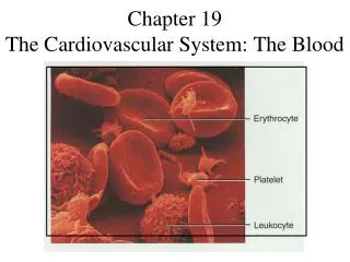
Figure 17.2: Photomicrograph of a human blood smear stained with Wright’s stain, p. 649. Platelets Erythrocytes Monocyte Neutrophils Lymphocyte
Figure 17.9: Types and relative percentages of leukocytes in normal blood, p. 657. Differential WBC count (All total 4800– 10,800/ml) Formed elements Granulocytes • Neutrophils (50–70%) Platelets Leukocytes • Eosinophils (2–4%) • Basophils (0.5–1%) Erythrocytes Agranulocytes • Lymphocytes (25–45%) • Monocytes (3–8%)
John Jacob Abel 1857 – 1938 Endocrinologist who was extensively involved with work on insulin and adrenalin. His most famous work however was in designing blood dialysis.
Figure 17.4: Structure of hemoglobin, p. 651. b1 2 H3C CH2CH2COOH N CH2CH2COOH H2C=CH N Fe N CH3 H3C N H2C=CH CH3 Polypeptide chain 2 a1 (b) Iron-containing heme group (a) Hemoglobin
Figure 17.6: Erythropoietin mechanism for regulating erythropoiesis, p. 653. Imbalance Start Homeostasis: Normal blood oxygen levels Stimulus: Hypoxia due to decreased RBC count, decreased amount of hemoglobin, or decreased availability of O2 Imbalance Increases O2- carrying ability of blood Reduces O2 levels in blood Kidney (and liver to a smaller extent) releases erythropoietin Enhanced erythropoiesis increases RBC count Erythropoietin stimulates red bone marrow
Figure 17.7: Life cycle of red blood cells, p. 654. Low O2 levels in blood stimulate kidneys to produce erythropoietin. 1 Erythropoietin levels rise in blood. 2 Erythropoietin and necessary raw materials in blood promote erythropoiesis in red bone marrow. 3 New erythrocytes enter bloodstream; function about 120 days. 4 Aged and damaged red blood cells are engulfed by macrophages of liver, spleen, and bone marrow; the hemoglobin is broken down. 5 Hemoglobin Heme Globin Bilirubin Iron stored as ferritin, hemosiderin Amino acids Iron is bound to transferrin and released to blood from liver as needed for erythropoiesis Bilirubin is picked up from blood by liver, secreted into intestine in bile, metabolized to stercobilin by bacteria and excreted in feces Circulation Food nutrients, including amino acids, Fe, B12, and folic acid are absorbed from intestine and enter blood Raw materials are made available in blood for erythrocyte synthesis. 6
Figure 17.5: Erythropoiesis: genesis of red blood cells, p. 652. Stem cell Committed cell Developmental pathway Phase 1 Ribosome synthesis Phase 2 Hemoglobin accumulation Phase 3 Ejection of nucleus Early erythroblast Late erythroblast Hemocytoblast Proerythroblast Normoblast Reticulocyte Erythrocyte
Figure 17.11: Leukocyte formation, p. 661. Hemocytoblast Stem cells Lymphoid stem cell Myeloid stem cell Myeloblast Myeloblast Myeloblast Lymphoblast Committed cells Promyelocyte Promyelocyte Promyelocyte Promonocyte Prolymphocyte Develop- mental pathway Eosinophilic myelocyte Basophilic myelocyte Neutrophilic myelocyte Eosinophilic band cells Neutrophilic band cells Basophilic band cells Monocytes Eosinophils Basophils Neutrophils Lymphocytes (a) (b) (c) (d) (e) Agranular leukocytes Some become Granular leukocytes Some become Plasma cells Macrophages (tissues)
Figure 17.12: Genesis of platelets, p. 662. Stem cell Developmental pathway Hemocytoblast Megakaryoblast Promegakaryocyte Megakaryocyte Platelets
Figure 17.15: Blood typing of ABO blood types, p. 671. Blood being tested Serum Anti-A Anti-B Type AB (contains agglutinogens A and B) RBCs Type B (contains agglutinogen B) Type A (contains agglutinogen A) Type O (contains no agglutinogens)