Download
1 / 36
360 likes | 533 Views
Physical Examination: Neurological. Nose Exam. Smell test CN I. Patient closes eyes and plugs one nostril. Hold an alcohol swab a few inches away and have them sniff. Repeat with the other nostril. Is the strength of the smell the same?. Eye Exam: testing cranial nerves III, IV, and VI.

E N D
Nose Exam • Smell test • CN I Patient closes eyes and plugs one nostril. Hold an alcohol swab a few inches away and have them sniff. Repeat with the other nostril. Is the strength of the smell the same?
Eye Exam: testing cranial nerves III, IV, and VI • Instruct the patient to hold his head still and follow the examiner’s finger with his eyes as the examiner circumscribes a large "H" in front of the patient. Then move the finger towards the patient’s nose. Weakness in an eye muscle indicates cranial nerve damage. • CN III also controls the pupillary reflex. Have the patient look straight ahead, then shine a light into the left eye once to see if the left pupil constricts. Remove the light and shine it again at the left eye, but look at the right eye to see if the right pupil constricts. • Repeat this, shining the light twice in the right eye to see if the right and left pupil constrict.
Look for Nystagmus • Nystagmus is involuntary eye movement. • http://en.wikipedia.org/wiki/Physiologic_nystagmus • It is characterized by alternating smooth pursuit in one direction and saccadic movement in the other direction. • Pathological nystagmus is the result of damage to one or more components of the vestibular system, including the semicircular canals, otoliths, and the vestibulocerebellum. • Many blind people have nystagmus, which is one reason that some wear dark glasses.
Extraocular Movements: CN III (except as listed) CN VI CN IV
Retinal Exam • Have the patient stare at a the corner of the room where the ceiling meets the wall. • Approach the right eye from the right side of the patient. Place one hand on their forehead so you have depth perception and don’t run into their face. • Place the ophthalmoscope on your forehead and look into the pupil. Examine the fundus (interior of the eye) for abnormalities.
Hearing Loss • Conductive hearing loss happens when there is a problem conducting sound waves through the outer ear, tympanic membrane (eardrum) or middle ear (ossicles). • Sensorineural hearing loss is a problem in the vestibulocochlear nerve (Cranial nerve VIII), the inner ear, or central processing centers of the brain.
Weber Test • Weber Test: only tests unilateral problems. A tuning fork is touched to the middle of the forehead: • Sensorineural hearing loss: sound is heard louder in the normal ear because the damage is to the nerve, so bone conduction of the sound is ineffective. • Conductive hearing loss: sound is heard louder in the problem ear (earwax, etc) because reflected soundwaves cannot escape the ear canal, so they penetrate deeper into the inner ear.
Rinne Test • Performed by placing a vibrating tuning fork on the mastoid process until sound is no longer heard, the fork is then immediately placed just outside the ear. Normally, the sound is audible at the ear, indicating a positive Rinne test. • If they cannot hear the sound at the ear, it is a negative Rinne test, and indicates Sensorineural hearing loss
Middle Ear Exam • Make sure a disposable specula is on the otoscope! • Approach the patient from the side. • Grasp the pinna (external ear) and gently pull it superiorly and posteriorly to straighten out the ear canal. • Place the specula of the otoscope into the ear first, before looking into the otoscope. Otherwise, you might drive the specula in too deep and hurt the patient. • Then look into the otoscope. Examine the canal for redness, look for perforation or scars on the tympanum (ear drum).
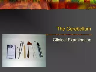
Gait and Station • Tandem gait • walk a straight line while touching the heel of one foot to the toe of the other with each step. Tests cerebellar function. • Hopping on one foot (not for feeble patients) • Romberg • Ask the patient to stand with their feet together (touching each other). Then ask the patient to close their eyes. Remain close in case the patient begins to sway or fall. • With the eyes open, visual input helps to maintain stability. Closing the eyes and having difficulty with the task suggests a mild lesion in the vestibular system.
Cerebellar Function • Finger to Nose • Patient is asked to alternately touch their nose and the examiner's finger as quickly as possible, while the examiner's finger is held at the extreme of the patient's reach, and the examiner's finger is occasionally moved • Heel to Shin • Touch the heel of one foot to the opposite knee and then to drag their heel in a straight line all the way down the front of their shin and back up again. In order to eliminate the effect of gravity in moving the heel down the shin, this test should always be done in the supine position. • Rapid Alternating Movements • Wiping one palm alternately with the palm and dorsum of the other hand
Reflexes • Biceps (C5-6) • Triceps (C7) • Patellar (L4) • Ankle (S1) • Plantar • Babinski sign 0: absent reflex 1+: trace 2+: normal 3+: brisk 4+: nonsustained clonus (i.e., repetitive vibratory movements) 5+: sustained clonus Deep tendon reflexes are normal if they are 1+, 2+, or 3+ unless they are asymmetric or there is a dramatic difference between the arms and the legs. http://www.neuroexam.com/content.php?p=2 http://meded.ucsd.edu/clinicalmed/neuro3.htm
Reflexes Exam • The limbs should be in a relaxed and symmetric position. • The muscle group to be tested must be in a neutral position (i.e. neither stretched nor contracted). • Compare each reflex immediately with its contralateral counterpart so that any asymmetries can be detected. • If you cannot elicit a reflex, you can sometimes bring it out by certain reinforcement procedures. For example, have the forcefully contract a different muscle group when the reflex is tested.
Biceps Reflex • Have the patient’s elbow resting in your arm at a 90 degree angle. • Place your thumb over the antecubitalfossa (inside of elbow) of the patient. • Ask the patient to flex their forearm (i.e. contract their Biceps muscle) while you simultaneously palpate the fossa. The Biceps tendon should become taut and thus readily apparent. • Place your thumb over the tendon and tap the pointed side of the reflex hammer onto your thumb and observe the reflex in their biceps.
Triceps Reflex • Have the patient’s elbow resting in your arm at a 90 degree angle. • Ask the patient to extend their forearm (i.e. contract their Triceps muscle) while you simultaneously palpate the tendon behind the elbow. • Place your thumb over the tendon and tap the pointed side of the reflex hammer onto your thumb and observe the reflex in their triceps.
Patellar Reflex • Make sure the leg is relaxed. • Use the wide side of the hammer. • If no reaction, have patient clasp fingers together and pull while the test is performed.
Achilles Tendon Reflex • This is most easily done with the patient seated, feet dangling over the edge of the exam table. • Use one hand to hold the foot at a 90 degree angle, and strike the tendon with the wide side of the hammer in the other hand. Feel for the foot plantarflexion.
Plantar Reflex • Scrape an object across the sole of the foot beginning from the heel, moving forward toward the small toe, and then arcing medially toward the big toe. • The normal response is downward contraction of the toes. • The abnormal response, called a positive Babinski's sign, is characterized by a dorsiflexed big toe and fanning outward of the other toes. This is normal in children under 1 year of age, but abnormal in adults.
Cranial Nerves • Olfactory (I) smell • Optic (II) Visual Acuity, • Pupil Reactions, • Extraocular Movement (III) • Nystagmus • Superior oblique (Trochlear IV) • Lateral Rectus (Abducins VI) • Trigeminal (V) sensory of face • Facial Motor (VII) blink and smile • Vestibulocochlear (VIII) hearing/balance • Glossopharyngeal (IX) swallowing • Vagus (X) larynx moves with speech • Accessory (XI) shrug shoulders • Hypoglossus (XII) move tongue
CN V • Check sensation on the upper, middle, and lower part of the face. • Check for ability to open and close mouth in a chewing fashion.
CN VII • Voluntary facial movements, such as wrinkling the brow, showing teeth, frowning, closing the eyes tightly, pursing the lips and puffing out the cheeks, all test the facial nerve. There should be no noticeable asymmetry. • CN VII also supplies taste to the anterior 1/3 of tongue.
Sensation • Hot, Cold • Pain, Light Touch • Vibration • Proprioception • Two-point discrimination Perform each of these tests throughout the dermatome map.
Map of Dermatomes Test from the sides of the neck , to the shoulder Circumscribe the upper and lower arms Test down the front of the chest or back Test the lateral sides of the thigh Circumscribe the leg
Sensation • Temperature sensation can be tested with a cool piece of metal such as a tuning fork or stethoscope diaphragm. Warms can be tested with warm hands or warmed cloth. • Light touch is best tested with a cotton-tipped swab • Pain is tested by poking gently with a sharp object, such as a broken wooden stick from a Q-Tip
Sensation • Vibration • Place a vibrating tuning fork on the fleshy portion of the patient's toe or finger and ask him to report when the vibration stops. • Take care not to place the tuning fork on a bone, since bones conduct the vibration to much more proximal sites, where they can be detected by nerves far from the location being tested.
Sensation • Proprioception (joint position) • Move one of the patient's fingers or toes up and down and asking the patient to report which way it moves. • Hold the digit lightly by the sides while doing this so that tactile inputs don't provide significant clues to the direction of movement. • The digit should be moved very slightly because normal individuals can detect movements that are barely perceptible by eye.
Sensation • Two-point discrimination • Use a caliper or bent paper clip, alternating randomly between touching the patient with one or both points. • Ask them each time: can you feel one prong or two? If you use two and they say “one”, spread the clip points wider and try again. • The minimal separation (in millimeters) at which the patient can distinguish these stimuli should be recorded in each extremity.
Muscle Strength • Upper Extremity • Shoulder • Elbow • Wrist • Lower Extremity • Hip • Knee • Foot 0/5: no contraction 1/5: muscle flicker, but no movement 2/5: movement possible, but not against gravity (test the joint in its horizontal plane) 3/5: movement possible against gravity, but not against resistance by the examiner 4/5: movement possible against some resistance by the examiner 5/5: normal strength
Muscle Strength • Pair the testing of each muscle group immediately with testing of its contralateral counterpart to enhance detection of any asymmetries. • Have them flex, then extend, the chosen joint. First the right, then the left. • Have them flex and extend that joint again, but this time, examiner pushes against the movement, creating resistance. • Proceed to the next joint.