Download
1 / 33
360 likes | 944 Views
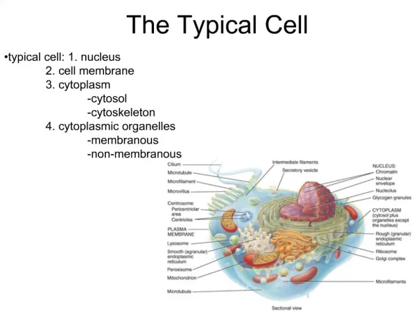
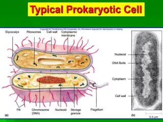
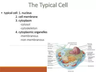
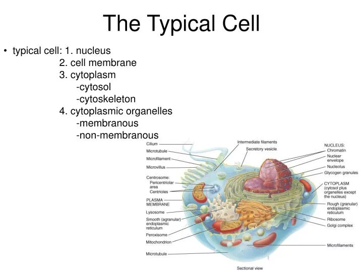
The Typical Cell. typical cell: 1. nucleus 2. cell membrane 3. cytoplasm -cytosol -cytoskeleton 4. cytoplasmic organelles -membranous -non-membranous. Cytosol. ECF. lower K+ higher Na+ lower concentration of dissolved and suspended proteins

E N D
The Typical Cell • typical cell: 1. nucleus • 2. cell membrane • 3. cytoplasm • -cytosol • -cytoskeleton • 4. cytoplasmic organelles • -membranous • -non-membranous
Cytosol ECF • lower K+ • higher Na+ • lower concentration • of dissolved and • suspended proteins • higher concentration • of carbohydrates • smaller reserves of amino • acids • higher K+ • lower Na+ • higher concentration • of dissolved and • suspended proteins • (enzymes, organelles) • lower concentration • of carbohydrates • (due to catabolism) • larger reserves of amino • acids (anabolism) • Cytoplasm • semi-fluid-like jelly within the cell • division into two subdivisions: cytosol & organelles • also contains a supportive framework of proteins: cytoskeleton Cytosol -intracellular fluid of the cell -about 55% of the cell’s volume -about 70-90% water PLUS dissolved nutrients ions soluble & insoluble proteins waste products glucose, ATP amino acids, fatty acids
Cytoskeleton: • internal framework of the cell • gives the cytoplasm flexibility and strength • three major components • 1. microfilaments • 2. intermediate filaments • 3. microtubules
microfilaments = made of thin filaments called actin -forms a dense network immediately under the PM + scattered throughout the cytoplasm (most prevalant at the cell periphery) -function: 1. anchor integral proteins and attaches them to the cytoplasm 2. interacts with larger filaments made up of myosin - results in active movements of the cell (e.g. muscle cells) or changes in cell shape -provide much of the mechanical strength of the cell + give the cell its shape -also provide support for cellular extensions called microvilli (small intestines)
3. Type III: vimentin (bone, cartilage) -desmin (muscle) intermediate filaments = made up of vimentin, desmin or keratin -function: 1. impart strength 2. stabilize organelles 3. transport materials -some IFs are made of specialized proteins found in specific cell types e.g. neurofilaments in neurons - for transport of synaptic vesicles containing neurotransmitters • five major groups of IFs 1. Type I: acidic keratins (epithelial cells) 2. Type II: basic keratins (epithelial cells) 4. Type IV: Neurofilaments (neuronal) 5. Type V: Lamins A, B, C (all cells)
microtubules= repeating units of tubulin -assembly is controlled in the MTOC (microtubule organizing center) - located near the nucleus – region of tubulin proteins -for the assembly of tubulin into microtubules, centrioles and cilia -also known as a centrosome -role in MT assembly -found near the Golgi apparatus during interphase - called the centrosome (pair of centrioles at right angles) -MTs grow out from the centrosome -microtubule – a cylinder = 13 rows of tubulin arranged as a “straw” -straws can be glued together to form a doublet or a triplet -function: 1. cell shape & strength 2. organelles: anchor & movement 3. mitosis - form the spindle (chromosome movement) 4. form many of the non-membranous organelles (cilia, flagella, centrioles)
Non-membranous Organelles A. Centrioles:short cylinders of tubulin – arranged as 9 short microtubule triplets -called a 9+0 array (9 peripheral triplets, 0 in the center) -each centriole is made of a pair – arranged perpendicular to one another -role in mitosis - spindle and chromosome alignment
cilia 9+2 basal body 9+0 B. Cilia & Flagella • cilia = contain 9 groups of microtubule doublets surrounding a central pair • -called a 9+2 array • -anchored to a basal body just beneath the cell surface (9+0 array) • -exposed portion of the cilia covered by PM – but the cilia itself is non-membranous • -beat rhythmically to transport material • -found in linings of several major organs covered with mucus • -function in “cleaning”
flagella = resemble cilia • -much larger • -found singly • -functions to move a cell through the ECF • ??? What is the only human cell to possess a flagella???
Ribosomes = can be considered a nonmembranous organelle • 2 protein subunits in combination with RNA • -large 60S subunit = 28S rRNA (ribosomal RNA) + 50 proteins • -small 40S subunit = 18S rRNA + 33 proteins • actual site of mRNA translation -> peptide strand • in association with the ER = where the peptide strand • is fed into from the ribosome • also float freely within the cytoplasm as groups = polyribosomes
Membranous Organelles • completely surrounded by a phospholipid bilayer similar to the PM • surrounding the cell • allows for isolation of each individual organelle - so that the components • of each organelle does not mix with the cytosol • therefore requires a well-coordinated system of transport for organelles • to communicate and function together • -”vesicular transport” - small transport vesicles pinch off one • organelle, travel and then fuse to another organelle
1. Endoplasmic reticulum (ER) = series of membrane-bound, flattened sacs in communication with the nucleus and the PM -each sac or layer = cisternae -inside or each sac =lumen -two types: Rough ER - outside studded with ribosomes -continuous with the nuclear membrane -protein synthesis, phospholipid synthesis -the peptide strand as it is being translated by the ribosome is fed into the RER -initial site of processing (attachment of carbohydrates) and sorting for transport to the Golgi -three functions: 1. synthesis 2. storage 3. transport
transport from the ribosome across the ER membrane requires the presence of a signal sequence • 16-30 amino acids at the beginning of the peptide sequence (N-terminal) • this signal sequence will vary from protein to protein by will have a few characteristics in common – starts with one or two positively charges amino acids and is followed by 6-12 hydrophobic AAs • A complex of proteins will bind this signal in the cytoplasm = signal recognition particle/SRP • the ER membrane has receptor for the SRP – docks the translating ribosome (yellow protein in figure) • actual entrance of the protein requires the formation of a “hole” in the ER membrane (blue protein in figure) = translocon (series of multiple transmembrane proteins) • this process also requires energy which is provided by GTP
Translocation • this animation might be a bit complicated – but give it a try • http://www.rockefeller.edu/pubinfo/proteintarget.html
Modifications in the RER • 1. formation of disulfide bonds • help stabilize the tertiary and quaternary structure of proteins • 2. folding of the peptide chain • misfolded proteins remain in the cytoplasm and are degraded • only properly folded proteins get transported to the Golgi for additional processing and transport • many proteins located in the ER which stimulate this folding • 3. addition and processing of carbohydrates • 4. breakage of specific peptide bonds – proteolytic cleavage • 5. assembly into multimeric proteins (more than one chain) for an animation go to http://sumanasinc.com/webcontent/animations/content/proteinsecretion_mb.html
Smooth ER – extends from the RER but is free of ribosomes • many enzymes on the surface of the SER for: • - 1. lipid and steroid (e.g. estrogen and testosterone) biosynthesis for membranes • - 2. detoxification of toxins and drugs • 3. vesicle formation • 4. cleaves glucose so it can be released into the bloodstream • -the amount of SER per cell can increase with drug use – cell accomadatesto increase protection
3. Golgi Apparatus = stack of 3 to 20 flattened membrane sacs/cisternae • site of protein modification, and final packaging of the • finished protein into secretory vesicles -> exocytosis or for use in the cytosol • movement of protein through the stacks via transport vesicles • -definite direction: first stack = cis-face (from the RER, protein modification) • middle stack = medial-face (adds carbohydrates) • last stack = trans-face (modification and packaging into vesicles)
Modifications in the Golgi • glycosylation = glycoprotein • most plasma membrane and secreted proteins have one or more carbohydrate chains that help target them to the correct location • some glycosylation occurs in the ER, others in the various sacs of the Golgi • in the Golgi are the addition of N- and O-linked oligosaccharides – specific sugar residues • O-linked are added one at a time in the Golgi to the amino acids serine, threonine or lysine (one to four saccharide subunits total) • added on by enzymes called glycosyltransferases • human A, B and O antigens are sugars added onto proteins and lipids - inserted in the PM of the RBC • everyone has the glycosyltransferase needed to produce the O antigen • N-linked saccharides are linked together (about 14 sugars!) in the ER • transported out of the ER and imported into the Golgi • are attached specifically to asparagine amino acids within a protein • specific sugars can be removed or new ones added in the Golgi – changes the composition of the resulting glycoprotein • the sugars are found in the cytoplasm but are transported into the Golgi by specific transporters for their addition
Modifications in the Golgi • some PM proteins and most secretory proteins are synthesized as larger, inactive proproteins that will require additional processing to become active • e.g. albumin, insulin, glucagon • this processing occurs very late in maturation = trans face • processing is catalyzed by protein-specific enzymes called proteases • some proteases are unique to the specific secretory protein • occurs in secretory vesicles that bud from the trans-Golgi face • processing could be at one site (albumin) others may require more than one peptide bond (insulin)
-proteins processed in the Golgi will have unique sequences of amino acids that tell the protein where to go -these are called sorting signals -the lack of this signal means you will be secreted • targets: • secretory vesicles for exocytosis • membrane vesicles for incorporation into PM • transport vesicles for intracellular destinations • e.g. digestive enzymes to the lysosome • -requires unique sorting • signals
4. Mitochondria = site of energy production (ATP production) -via Cellular Respiration - breakdown of glucose into water and CO2 results in the production of ATP -initial glucose breakdown occurs in the cytosol -terminal stages occur in the mitochondria = Oxidative Phosphorylation -has its own DNA - maternal -reproduce themselves via dividing -consists of an outer mitochondrial membrane, an inner mitochondrial membrane and a fluid-filled space = mitochondrial matrix (contains ribosomes!) -the inner membrane is folded into folds called cristae (increase the membrane surface area for the enzymes of Oxidative Phosphorylation)
outer - 50% lipid & 50% protein • -very permeable - contains pores -2 membrane layers: • inner - 20% lipid & 80% protein • -less permeable • -folded extensively to form partitions = cristae • -contains proteins that transport H+ out of the • lumen of the mitochondria -> electrochemical gradient • -contains enzymes that use this gradient for the synthesis • of ATP • -also contains pumps to move ATP into the cytosol • matrix - lumen of the mitochondria • -breakdown of glucose into water and CO2 ends here and results in the production of ATP
Cellular respiration -glycolysis -citric acid cycle -electron transport chain http://biology.about.com/gi/dynamic/offsite.htm?site=http://www.sp.uconn.edu/%7Eterry/images/anim/ETS.html http://biology.about.com/gi/dynamic/offsite.htm?site=http://www.biocarta.com/pathfiles/krebPathway.asp http://vcell.ndsu.nodak.edu/animations/etc/movie.htm
Glycolysis phosphorylation • literally means “splitting sugar” • conversion of glucose (6 carbon sugar) into 2 molecules of pyruvate(3 carbon sugar) • pyruvate will be converted into acetyl-coenzyme A (Acetyl-CoA) which then enters the citric acid cycle • under aerobic conditions glucose is oxidized into pyruvate and then continues on to the citric acid cycle • under anaerobic conditions glucose is oxidized into pyruvate then converted into lactate (lactic acid) • reactions of glycolysistake place in the cytosol • this pathway also creates the building blocks that are used for the synthesis of long chain fatty acids isomerization phosphorylation cleavage 2 ATP consumed no energy created key enzyme in controlling the rate of glycolysis = phosphofructokinase -liver enzyme that is inhibited when ATP levels are high http://web.indstate.edu/thcme/mwking/glycolysis.html http://science.nhmccd.edu/biol/glylysis/glylysis.html
Glycolysis • we consume sugars other than glucose • two other abundant sugars are fructose and galactose • fructose can be converted into glyceraldehyde using the fructose-1-phosphate pathway which utilizes different enzymes but still creates glyceraldehyde 3-phosphate • alternatively fructose can be phosphorylated to fructose-6-phosphate by the same enzyme that phosphorylates glucose (hexokinase) – then is phosphorylated again = fructose-1,6-phosphate • galactose can be converted into glucose-6-phosphate in a series of four steps using four different enzymes
Glycolysis • the cells in all kinds of organisms convert glucose to pyruvate using similar reactions • however pyruvate can be processed in many ways to produce • 1. ethanol – by yeast • pyruvate – acetylaldehyde – ethanol • regulated by aldehyde dehydrogenase (DH = removes H+ from one substrate and adds it to another) • the opposite (alcohol to aldehyde) is catalyzed by alcohol dehydrogenase (ADH) • 2. lactate – by cells in the absence of oxygen • 3. acetyl CoA – the majority of pyruvate is processed by this next phase
Citric Acid cycle • pyruvate is transported from the cytosol across the membranes of the mitochondria into its matrix • interacts with coenzyme A (mitochondrial enzyme) to produce acetyl-CoA • forms the waste product carbon dioxide (2 molecules) • Acetyl-CoA is converted into oxaloacetic acid -> citric acid • the citric acid is converted into a series of compounds that eventually regenerates OA acid • takes place in the matrix and inner membrane of the mitochondria • cycle results in the formation of NAD+ (nicotinamide adenine dinucleotide) – capable of storing high energy electrons • as the cycle runs - NAD+ is reduced to form NADH and H+ (gains two electrons) • NADH = electron carrier • NAD+ is capable of adding one H+ and 2 electrons • GTP hydrolysis required. • this NADH will then enter the electron transport chain • Also generates the FADH2 electron carrier (for 4 electrons) • while this cycle only runs in the presence of oxygen – no oxygen is used For each glucose this cycle has to “turn” Twice!!
Electron transport chain cytochrome c reductase cytochrome c oxidase NADH dehydrogenase • protons are pumped from the matrix into the space between the inner and outer mitochondrial membranes by enzyme complexes • in addition, electrons are transferred from these complexes to oxygen eventually forming water and carbon dioxide • protons are taken from NADH and pumped across the inner mitochondrial membrane by NADH dehydrogenase • at the same time electrons are moved from NADH dehydrogenase to cytochrome c reductase (by Q = ubiquinone) • cytochrome c reductase pumps more protons (from water) across the membrane and more electrons are transferred to cytochrome c oxidase (by cytochrome c) • cytochrome c oxidase also pumps more protons (taken from water) • this creates a proton gradient = energy • as protons flow down their gradient back into the matrix, they pass through an enzyme called ATP synthase – which synthesizes ATP protons electrons http://biology.about.com/gi/dynamic/offsite.htm?site=http://www.sp.uconn.edu/%7Eterry/images/anim/ETS.html
ETC animations • http://www.youtube.com/watch?v=xbJ0nbzt5Kw&feature=relmfu • http://www.youtube.com/watch?v=3y1dO4nNaKY • http://biology.about.com/gi/dynamic/offsite.htm?site=http://www.sp.uconn.edu/%7Eterry/images/anim/ETS.html
5. Lysosomes= “garbage disposals” -dismantle debris, eat foreign invaders/viruses taken in by endocytosis or phagocytosis -also destroy worn cellular parts from the cell itself and recycles the usable components = autophagy -form by budding off the Golgi -vesicle sacs that contain powerful enzymes to breakdown substances into their component parts e.g. nucleases = breakdown RNA & DNA into nucleotides proteases = breakdown proteins into amino acids -over 60 kinds of enzymes within the lysosome -these enzymes are collectively known as acid hydrolases -acidic interior - critical for function of these enzymes -created and maintained by a hydrogen pump that transport H+ into the interior - Active transport -chloride ions that diffuse in passively through a chloride channel - forms hydrochloric acid (HCl)
6. Peroxisomes: abundant in liver and kidney cells -main function is the breakdown of long chain fatty acids through their oxidation -oxidation is done by oxidases = enzymes that use oxygen to oxidize substances - such as peroxidase -oxidation reactions by these oxidases generate toxic oxidative chemicals such as hydrogen peroxide (H2O2) -therefore peroxisomes contain enzymes to break up these oxidative substances e.g. catalase to break this peroxide down - catalyze the breakdown of H2O2 into water and oxygen -outer membrane contains a variety of enzymes for 1. synthesis of bile acids 2. breakdown of lipids - oxidation of fatty acids and amino acids (formed during normal metabolic processes) 3. detoxification of alcohol – converts it into sugars F-actin and peroxisomes
Diseases at the Organelle Level 1. Tay Sachs and lysosomes: lack one of the 40 lysosomal enzymes -produces a lysosomal storage disease -> build up of fatty material on nerve cells -failure of nervous system communication 2. Adrenoleukodystrophy and peroxisomes: peroxisomes lack an enzyme on the outer membrane which transports an essential enzyme into the peroxisome -leads to a build up of a long-chain fatty acid on cells of the brain and spinal cord -> loss of the myelin sheath -lethargy, skin darkens, blood sugar drops, altered heart rhythm imbalanced electrolytes, paralysis, death *** slowed by a certain triglyceride found in rapeseed oil Lorenzo Odone = “Lorenzo’s Oil”