Download
1 / 13
140 likes | 254 Views
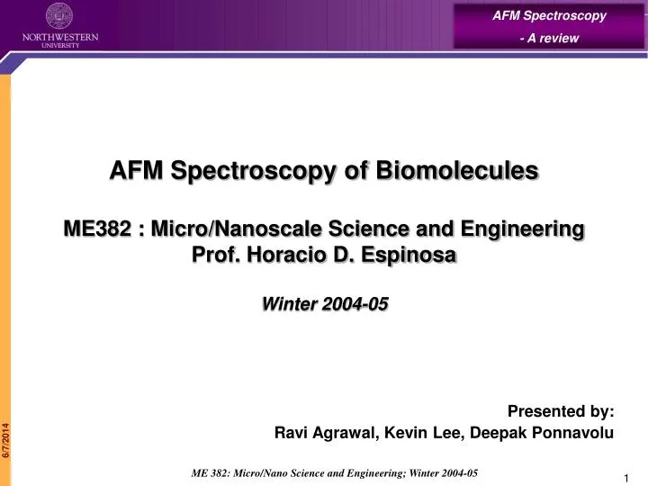
AFM Spectroscopy of Biomolecules ME382 : Micro/Nanoscale Science and Engineering Prof. Horacio D. Espinosa Winter 2004-05. Presented by: Ravi Agrawal, Kevin Lee, Deepak Ponnavolu. Outline. Introduction and Working Principle AFM in liquids Substrate Functionalization

E N D
AFM Spectroscopy of BiomoleculesME382 : Micro/Nanoscale Science and EngineeringProf. Horacio D. EspinosaWinter 2004-05 Presented by: Ravi Agrawal, Kevin Lee, Deepak Ponnavolu
Outline • Introduction and Working Principle • AFM in liquids • Substrate Functionalization • Tip Functionalization and Tip parameters • Developments and Applications • Typical force curve • Conclusion
Tip – Sample Interaction Schematic diagram of the vertical tip movement during the approach and retract parts of a force spectroscopy experiment* Tip Far Away (10-100 microns) No interactions Tip Approaching (few microns) Electrostatic Forces Tip Close to Surface Van der Waal Forces Capillary Forces Contact Tip Indenting the sample Stiffness Viscoelastic Response Lifting Off Surface (few atomic distances to nanometers) Tip farther away (nanometers to hundreds of nanometers) Stretched molecules between tip and surface Protein unfolding, pulling out of membranes Tip Far Away (1-5 microns) Connection between the tip and substrate is broken. No more interactions *Technical Report Nanowizard – A practical guide to AFM force spectroscopy and data analysis
Working Principle • Linker molecules are used to functionalize the tip and the substrate • Ligand and receptor molecules are attached to the linker molecules • The functionalized substrate holding the receptor molecule is approached with AFM tip • Ligand recognizes the receptor and fits in • AFM tip can be retracted to study the response of the ligand-receptor molecule
AFM in Liquids – Biological Applications • Why in Liquid ? • To prevent contamination of sample from hydrocarbons present in air • Most of the biomolecules are active in aqueous environment • Most of the cells (except few epithelial ones) die in dry conditions • AFM in liquids • Higher amplitude of oscillation to get good signal to noise ratio • Quality factor (~wom/b) goes down drastically because of high hydrodynamic damping • Typical thickness of cantilevers in tapping mode ~ 10 microns. By increasing this thickness, Q-factor as well as stiffness can be increased to an optimum value • Included angle of the tip should be lower in order to cut down the van der Waals forces
Substrate Functionalization • Why do we need to functionalize the substrate ? • To hold the molecules onto the substrate • To align the molecules in a particular way to maintain selectivity DNA motion during scanning (A–C). AFM images in TE buffer over a 300 × 300 nm area. The images in A–C were taken with an interval of 4.5 min* *http://www.pubmedcentral.gov/articlerender.fcgi?tool=pmcentrez&artid=19541
Substrate Functionalization • Binding of biological molecules to a solid substrate • poly-L-lysine or poly-L-arginine • silanizing a solid surface with 3-aminopropyltriethoxysilane (APTES) • Cross-linking group via the amino end of APTES on a glass surface (ANB-NOS). • ultraflat Au(111) surface is used as a substrate for N-hydroxysuccinimide terminated self-assembled monolayers • For protein adsorption, Interaction forces include dipole and induced dipole moments, hydrogen bond forces and electrostatic potentials Schematic representations of several proteins attached to the substrate that are exposed to mechanical stress* *Thomas E. Fisher, Mariano Carrion-Vazquez, Andres F. Oberhauser, Hongbin Li, Piotr E. Marszalek, Julio M. Fernandez. “Single Molecular Force Spectroscopy of Modular Proteins in the Nervous System” Neuron, Vol. 27, Sep. 2000, 435-446
Tip Functionalization • Why do we need to functionalize the tip? • To alter the surface properties of the tip so that it gains affinity to attach to the required end of the biomolecules • Helps in selectivity of single molecule Schematic Representation of Silanization Process* • Methods used for Functionalization: • Plasma Treatment to get desired hydrophobicity • Silanization • Using a spacer as a linker molecule in between – eg. PolyEthyleneGlycol (PEG) • Using appropriate biomolecules for linking – antigen or ligand Different ligands tethered to AFM tip via flexible PEG linker ** *C. Tolksdorf, I. Revenko “Choosing AFM Probes for Biological Applications” **Cordula M. Stroh et. al. “Tools for single molecule Recognition Force Microscopy and Spectroscopy” Molecular Imaging Application Note
Tip Parameters Tip Parameters play an important role for single-molecule recognition and spectroscopy • Main Features: • Tip Sharpness (Typically ~ 10 nm) • Dull tips for some biological applications – corrections required • Cantilever Stiffness – force measurements • Q-factor : typically 100~300 in air and 1 in liquid; using positive feedback system it can be controlled Sketch of a sensor molecule (brown) tethered to the AFM tip through a PEG linker * Sharpness of the tip required for biological applications ** *Cordula M. Stroh et. al. “Tools for single molecule Recognition Force Microscopy and Spectroscopy” Molecular Imaging Application Note ** http://www.lot-oriel.com/uk/htm/all/obe12b.php
Applications and Developments • 1) Cell Adhesion and Rupture • Contact Formation • Adhesion • Rupture Force Spectroscopy of adhesion between individual D. discoideum cells* • 2) Chemical Force Spectroscopy on Single-Walled Carbon Nanotubes • Nanotubes are good components for polymeric composites • Using Force Spectroscopy, their affinity to various molecules can be found 3) Estimating Bulk Properties using measurements at Molecular level *Martin Benoit, Daniela Gabriel, Gunther Gerisch, Hermann E. Gaub. “Discrete Interactions in cell adhesion measured by single-molecule force spectroscopy” Nature Cell Biology, Vol. 2, June 2000, 313-317
Typical Force Curve • The curve shows a typical force characteristics of a biomolecule (typically a folded protein): • Each drop in force (2-3) corresponds to one unfold in the molecular structure • Similar order of peaks show that same force is required to unfold it every time • Number of peaks tell about the total number of folds in the molecule The Forced extension of Modular Proteins exhibits a Saw-Tooth Pattern* *Thomas E. Fisher, Mariano Carrion-Vazquez, Andres F. Oberhauser, Hongbin Li, Piotr E. Marszalek, Julio M. Fernandez. “Single Molecular Force Spectroscopy of Modular Proteins in the Nervous System” Neuron, Vol. 27, Sep. 2000, 435-446
Conclusion • A very powerful tool for various applications like • Characterizing single biomolecules • Cell-cell interactions • Molecular interaction with surfaces • Challenges involved because of mechano-chemical interactions • Not very well-defined or specific process; Parameters of functionalization vary with different kind of molecules to be analyzed • Huge potential in upcoming era of Biotechnology
Thank You Questions ??


![Identification of Stereochemical Isomers of [Mo(CO) 4 (L) 2 ] by Infra-Red Spectroscopy](https://cdn2.slideserve.com/4500730/identification-of-stereochemical-isomers-of-mo-co-4-l-2-by-infra-red-spectroscopy-dt.jpg)