Download
1 / 133
1.33k likes | 1.46k Views
DO NOW:. Choose the part of your brain you would most hate to lose, and tell me why. Choose the part of your brain that you would like to enhance to give you a super power, and why. . Unit 3: Biological Bases of Behavior. AP Psychology Ms. Desgrosellier 10.28.2010. Neuropsychologists:.

E N D
DO NOW: • Choose the part of your brain you would most hate to lose, and tell me why. • Choose the part of your brain that you would like to enhance to give you a super power, and why.
Unit 3: Biological Bases of Behavior AP Psychology Ms. Desgrosellier 10.28.2010
Neuropsychologists: • psychologists who explore the relationships between brain/nervous systems and behavior. • aka: biological psychologists, biopsychologists, behavioral geneticists, physiological psychologists, and behavioral neuroscientists.
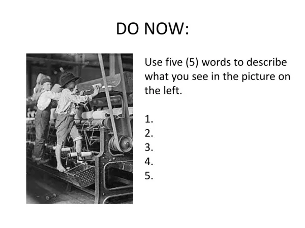
TECHNIQUES TO LEARN ABOUT STRUCTURE & FUNCTION • Clinical Observation (Case Study) • Look at injuries, diseases, etc.
TECHNIQUES TO LEARN ABOUT STRUCTURE & FUNCTION • Over 150 years ago people were studying patients with brain damage and linked loss of structure with loss of function. • Essentially losing brain tissue caused brain damage.
TECHNIQUES TO LEARN ABOUT STRUCTURE & FUNCTION • Phineas Gage was a level-headed, calm foreman of a railroad crew in 1848. • An explosion shot an iron rod through his head, severing the connections between his limbic system and his frontal cortex. • Gage became hostile, impulsive, and unable to control his emotions or his obscene language. • Autopsy revealed that the relationship between frontal lobes and control of emotional behavior.
Broca’s area • Paul Broca (1861) did an autopsy on a patient named Tan, who couldn’t speak even though there was no physical damage and he could understand language. • Tan’s brain showed loss of tissue in part of the frontal lobe of the left central cerebral hemisphere (as did several other similar cases).
Broca’s area • It was concluded that damage to this so-called Broca’s area caused a loss of ability to speak, known as expressive aphasia.
Wernicke’s area • Carl Wernicke found another brain area involved with understanding language in the left temporal lobe. • Destruction of Wernicke’s area results in loss of ability to comprehend written and spoken language, known as receptive aphasia.
DO NOW: • Briefly explain who Phineas Gage was and why he is important to Psychology.
Lesions • Precise destruction of brain tissue. • Enabled more systematic study of the loss of function resulting from surgical removal, cutting of neural connections, or destruction by chemical applications.
Lesions • E.g. Surgery to relieve epilepsy cuts neural connections at the corpus callosum, between cerebral hemispheres. • Studies of patients with “split brains” have shown that the left and right hemispheres do not perform exactly the same functions.
Right hemisphere: • nonverbal • spatial, musical, and holistic functions • identifying faces • recognizing emotional facial expressions
Left hemisphere: • verbal functions • mathematical functions • analytical functions • language
Manipulating the brain • Scientists can electrically, chemically, or magnetically stimulate various parts of the brain and note effects. • Researchers have electrically stimulated different cortical areas of the brain during surgery.
Manipulating the brain • It has enabled scientists to observe results, like: • the frontal cortex at particular sites caused body movement for different body parts enabling mapping of the motor cortex. • New research has found that you can magnetically lesion parts of the brain (temporary and so far has shown no harm)
DO NOW • Tell me at least three functions of the left hemisphere and three functions of the right hemisphere of the brain.
Brain Imaging • Computerized axial tomography (CAT or CT): two-dimensional x-ray slices that are passed through various angles of the brain, arranged to show the extent of a lesion.
Brain Imaging • magnetic resonance imaging (MRI): a technique that uses magnetic fields and radio waves to produce computer-generated images that distinguish among different types of soft tissue; allows us to see structures within the brain.
Brain Imaging • Putting one’s head into a strong magnetic field aligns the spinning atoms. • A pulse of a radio wave disorients the atoms briefly. • When the atoms return to their normal spin, they release signals that give us a detailed image of the body.
Measuring brain function • Scientists can stick a tiny microelectrode into a single neuron to measure its activity.
Measuring brain function • electroencephalogram (EEG): an amplified recording of the waves of electrical activity that sweep across the brain’s surface. These waves are measured by electrodes placed on the scalp.
Measuring brain function • The amplified tracings are called evoked potentials when the recorded changes in voltage results from a response to a specific stimulus presented to the subject. • Repeated study of the read-out can help researchers filter out brain activity and find the electrical wave caused by the specific stimulus.
Measuring brain function • functional magnetic resonance imaging (fMRI): a technique for revealing blood flow and, therefore, brain activity by comparing successive MRI scans. MRI scans show brain anatomy; fMRI scans show brain functions.
Measuring brain function • Researchers compare images taken less than a second apart, they can see which parts of the brain “light up” with increased blood flow.
Measuring brain function • positron emission tomography (PET) scan: a visual display of brain activity that detects where a radioactive form of glucose goes while the brain performs a given task.
Measuring brain function • Active neurons hog the glucose (the brain’s chemical fuel), and the PET scan tracks where in the brain the radioactive glucose goes.
Measuring brain function • Researchers can have participants think about certain topics or do activities to see where the glucose goes (thereby showing what part of the brain is active during that activity).
ORGANIZATION OF YOUR NERVOUS SYSTEM • All of the neurons in your body are organized into your nervous system. • The two major subdivisions are the central nervous system and the peripheral nervous system.
ORGANIZATION OF YOUR NERVOUS SYSTEM • Central Nervous System (CNS): made up of the brain and spinal cord. • Spinal cord: starts at the base of your back and extends upward to the base of your skull where it joins your brains. • Made mainly of interneuron’s and glial cells, which are all bathed by cerebrospinal fluid produced by your glial cells.
ORGANIZATION OF YOUR NERVOUS SYSTEM • Peripheral Nervous System (PNS): made up the somatic and autonomic nervous systems, and spread around your body from your spinal cord outwards. • Somatic Nervous System: motor neurons that stimulate skeletal (voluntary) muscle. • Autonomic Nervous System: motor neurons that stimulte smooth (involuntary) and heart muscle.
DO NOW • Describe one way of studying the brain and what it tells psychologists.
ORGANIZATION OF YOUR NERVOUS SYSTEM • The Autonomic Nervous System is divided into two parts: • Sympathetic Nervous System: Responses that help your body deal with stressful events, including: • Dilation of pupils, release of glucose from your liver, dilation of bronchi, inhibition of digestive functions, acceleration of heart rate, secretion of adrenalin from your adrenal glands, acceleration of breathing rate, and inhibition of secretion of your tear glands.
ORGANIZATION OF YOUR NERVOUS SYSTEM • The Autonomic Nervous System is divided into two parts: • Parasympathetic Nervous System: Calms your body following sympathetic stimulation by restoring digestive processes (salivation, peristalsis, enzyme secretion), returning pupils to normal size, stimulating tear glands, restoring normal bladder contractions, slow breathing and heart rate, etc.
ORGANIZATION OF YOUR NERVOUS SYSTEM • Turn to your neighbor and explain the two major subdivisions of the nervous system. • What are the 2 parts of the CNS? • What are the 2 parts of the PNS? • What are the 2 parts of the autonomic NS?
The Brain • Covered by protective tissue called meninges and housed in your skull. • The evolutionary perspective studies how the human brain has evolved. One theory breaks the brain into three sections: • The reptilian brain is similar to the brainstem in humans, and is responsible for maintaining homeostasis and instinctive behavior.
The Brain • The old mammalian brain roughly corresponds to the limbic system that controls emotional behavior, memory, and vision. • The new mammalian brain or cerebral cortex, accounts for 80% of the brain’s volume and is associated with higher functions of judgment, decision-making, abstract thought, foresight, hindsight, and insight.
The Brain • The surface of the cortex has peaks (gyri) and valleys (sulci), which form convolutions that increase the surface area of your cortex. • Deeper valleys are called fissures.
The Brain • The last evolutionary development of the brain is localization of functions on different sides of your brain.
LOCALIZATION AND LATERALIZATION OF THE BRAIN’S FUNCTION • Association areas: regions of the cerebral cortex that do not have specific sensory or motor functions, but are involved in higher mental functions, such as thinking, planning, remembering, and communicating.
LOCALIZATION AND LATERLIZATION OF THE BRAIN’S FUNCTION • Contralaterality: control of one side of your body by the opposite side of your brain. • The left side of your brain controls the right side of your body. • The right side of your brain controls the left side of your body.
DO NOW • Draw and label the Nervous System tree (diagram that separates the parts of the nervous system) Nervous System
Nervous System Peripheral Nervous System Central Nervous System Brain Spinal Cord Autonomic Nervous System Somatic Nervous System Sympathetic Nervous System Parasympathetic Nervous System
Structure of Brain: Brainstem • medulla: where most fibers cross above the brain stem, resulting in contralateral (opposite side) control. • regulates heart rate, blood flow, breathing, digestion, vomiting.
Structure of Brain: Brainstem • pons: right above the medulla, helps coordinate movement, and is the bridge between cerebral hemispheres and both medulla and cerebellum.
Structure of Brain: Brainstem • reticular formation: a nerve network in the brainstem (pons) that plays an important role in controlling arousal.
Structure of Brain • cerebellum: coordinates motor function integrating motion and positional information from the inner ear and muscles. • helps maintain balance.
Structure of Brain • basal ganglia (basal nuclei): links the thalamus with the motor cortex and other motor areas. • regulates initiation of movements, balance, eye movements, and posture. • Involved in reward/punishment learning and focus. • Some nuclei (neural clusters) involved in emotion.
Structure of Brain • thalamus: relay “station” for sensory pathways carrying visual, auditory, taste, and somatosensory information to/from appropriate areas of cerebral cortex. • Located at the top of the brain stem.