Download
1 / 39
400 likes | 441 Views
Explore the classification, anatomy, and functions of the nervous system, covering the central and peripheral components, as well as key structures like the brain, spinal cord, and meninges. Discover the roles of the autonomic nerve system, sensory organs, and the blood-brain barrier in maintaining homeostasis. Dive into the histology of brain regions like the cerebellum and cortex, understanding the intricate layers and cell types. Learn about reflex arcs and the unique features of the sense organs, including the ear's complex anatomy.

E N D
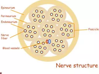
Nerve system 1. Classification of NS 2. CNS Brain: a) large hemispheres; b) cerebellum 3. Spinal cord 4. Meningeas 5. Blood-brain barrier 6. Peripheral nerve system. Spinal ganglia 7. Peripheral nerve 8. Nerve endings 9. Autonomic nerve system 10. Simple reflex arc Nervous system – special highly organized system (nervous tissue + connective)– intercommunicating network of neurons
CLASSIFICATION • Anatomical (structural): • central nervous system (CNS) – brain and spinal cord; • peripheral (PNS) – endings, fibers, ganglia, plexuses. • Functional: • a) somatic (voluntary, animal); • b) autonomic (involuntary, vegetative)
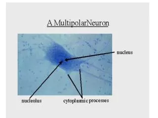
FUNCTIONS • 1. Integration • 2. Control • 3. Regulation • 4. Reception • 5. Conduction • 6. Analysis • 7. Response • NERVOUS SYSTEM ORIGIN • Ectoderm - nerve tube and ganglious lamella. • Cranial portion of nerve tube – brain and sense organs • Middle part of nerve tube and ganglious lamella– spinal cord, dorsal-root ganglia (spinal ganglia), autonomic ganglia and chromaffin tissue of human body. • Nervous tube zones • Ependymal – precursors of glial ependymal cells • Mantial layer – neuroblasts (nerve cells) and spongyoblasts (astrocytes and oligodendrocytes) • Marginal Zone – processes
BRAIN • Histologically:grey matter (nerve cells body) • white matter ( nerve fibers) • Grey matter: cortex + subcortical nuclei • MODUL – MFU of brain cortex • cilinder d 300 mkm around cortico-cortical fiber • NEUROPIL – aggregations of nerve and glial cells processes in central nerve system • Cytoarchitectonics - well regulated location of nervous cells (6 layers) • Myeloarchitectonics - well regulated location of nervous fibers (4 layers)
Cytoarchitectonics • Brain cortex has 6 layers • Pyramidal cells in the 2nd, 3rd, 4th, 5th layers • 1. Molecular • 2. Outer granular (10mkm) • 3. Pyramidal (10-40mkm) • 4. Inner granular • 5. Ganglionic (120x80, Betz, 1874) • 6. Multiform
MYELOARCHITECTONICS • 1. Above the 1st layer • 2. Under the 1st layer • 3. Above the 5th layer • 4. Under the 5th layer • TYPES OF NERVE FIBERS • Associative • Comissural • Projective
52 FIELDS OF BRODMANGRANULAR CORTEX – sensory (2nd , 4th)AGRANULAR CORTEX – motor (3rd,5th, 6)
CEREBELLUM Functions: 1. Coordination 2. Movement 3. Balance 4. Muscle tonus • Molecular layer: basket cells • large stellate cells • small stellate cells • Purkinje cells layer:Purkinje cells, supporting cells (lophogliocytes) • Granular layer:corn cells • stellate cells (2types) horizontal cells • Afferent fibers: • Mosslike– from olives and pons to the corn cells (tr. olivocerebellaris, tr. pontocerebellaris) • Climbing–from spinal cord and vestibular nuclei to the Purkinje cells (tr. spinocerebellaris, tr. vestibulocerebellaris) • Efferent fibers: axons of Purkinje cells
MENINGES • 3 protective coats of CNS: dura, arachnoid and pia mater • Skull bone • Periosteum of skull • epidural space • 1. DURA MATER– dense connective tissue • epithelium • subdural space • 2. ARACHNOID – flat epithelium • fibrocollagenous tissue • web-like strands • subarachnoid space • 3. PIA MATER – squamous epithelium • – loose connective tissue with blood vessels and nerve fibers • Basement membrane • Glia limitans (astrocytes) • Nerve tissue
BLOOD-BRAIN BARRIER • Prevents diffusion of substances from the blood to the brain • Capillary wall • 1. Endothelium • 2. Basement membrane • 3. Glial sheath (foot processes of astrocytes)
AUTONOMIC NERVE SYSTEM • Anatomically: a) central • b) peripheral • Functionally: a) sympathetic b) parasympathetic • SYMPATHETIC NS • Centers: thoracic-lumbar disposition • Nuclei intemediolateralis of spinal cord – multipolar associative radicular neurons • Sympathetic ganglia: paravertebral (trunci simpatici) and prevertebral (3) • PARASYMPATHETIC NS • Centers: cranio-sacral disposition • Nuclei of cranial nerves III, VII, IX and X pairs • Extramural and intramural ganglia (Dogel cells)
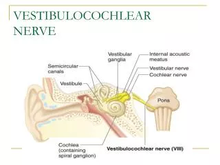
SENSE ORGANSEAR Taste buds Audiovestibular analizator Audiovestibular organ External ear Middle ear Internal ear: a) bony labyrinth; b) membranous labyrinth 7. Hair cells 8. Audiovestibular organ histophysiology
Organ of tasteneuroepithelial cells supporting cellsbasal cells
AUDIOVESTIBULAR ORGAN (EAR) External ear Middle ear Internal ear
INTERNAL EARvestibule+3 semicircular canals+cochlea Bony labyrinth Membranous labyrinth Otolith membrane Hair cells Maculae Cristae ampullaris Macula Organ of Corti Cristae ampullaris Cupula Hair cells Tectorial membrane Hair cells Crista ampullaris Organ of Corti
Cochlea axial section Helicotrema Scala vestibuli Modiolus Cochlear duct Spiral ganglion Scala tympani Cohlear nerve
MEMBRANOUS LABYRINTH • Cochlear duct
Membranous labyrinth (scheme) • Vestibular membrane • Basilar membrane • Stria vascularis Scala vestibuli Vestibular membrane Cochlear duct Spiral ligament Stria vascularis Tectorial membrane Spiral limbus Spiral tunnel Spiral bony lamella Basilar membrane Cochlear nerve Scala tympani
Corti’s organ Stria vascularis Hair cells Hensen cells Pillar cells Basilar membrane Outer phalangeal cells
Corti’s organ Outer cells: A. Supporting 1. Phalangeal (Deyters) 2. Border (Hensen) 3. Outer supporting (Claudius) B. Hair cells (3-5) Tunnel (pillar cells) Inner cells A. Supporting 1. Phalangeal 2. Inner supporting B. Hair cells (1-2) Outer hair cells Nerve fibers Tectorial membrane Spiral tunnel Inner hair cells Inner border cells Inner tunnel Cochlear nerve Outer border cells Outer supporting cells Inner phalangeal cells Betshar cells Inner and outer pillar cells Basilar membrane Outer phalangeal cells
Apical portion of outer phalangeal and hair cells 1 2 3 4 • Stereocilia • Cuticula • Phalangeal processes • Hair cells bodies
Phalangeal and hair cells (scheme) Stereocilia Cuticular lamella Marginal network Phalangeal process Outer hair cell Afferent and efferent nerve fibers Outer phalangeal cell Basilar membrane
Hair cells stereocilia Excitation Apical junctions Stereocilia Basal body Cuticular lamella Microtubuli
Audiovestibular organ histophysiology Subarachnoidal space Semicircular canals Ampulae Endolymphatic sac Endolymphatic duct Perilymphatic duct Scala vestibuli Cochlear duct Oval window Processsus mastoideus Scala tympani Stapes Uncus Maleus External auditory tube Cochlea Eustachian tube Tympanic membrane
Hearing histophysiology Scala vestibuli Oval window Vestibular membrane Cochlear duct Tectorial membrane Organ of Corti Round window Basilar membrane Cochlear nerve Scala tympani Spiral ganglion