Download
1 / 16
170 likes | 235 Views
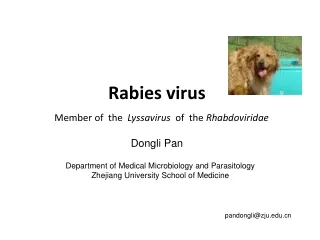
Rabies virus and Prion. Dongli Pan. Department of Medical Microbiology and Parasitology Zhejiang University School of Medicine. pandongli@zju.edu.cn. Rabies virus. M ember of the Lyssavirus of th e Rhabdoviridae. RNA viruses. DNA viruses. Baltimore classification.

E N D
Rabies virus and Prion Dongli Pan Department of Medical Microbiology and Parasitology Zhejiang University School of Medicine pandongli@zju.edu.cn
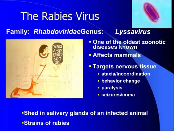
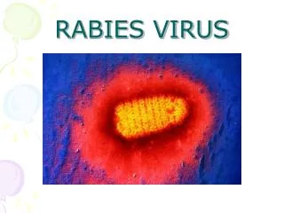
Rabies virus Member of the Lyssavirus of theRhabdoviridae
RNA viruses DNA viruses Baltimore classification Fields Virology, 6th edition
1. Biological properties Has a cylindrical morphology -ssRNA enveloped virus capsid has helical symmetry Is a neurotropic virus
Negri body 内基小体 Forms round or oval acidophilic cytoplasmic inclusion bodies. Negri body in the cytoplasm of infected neuron
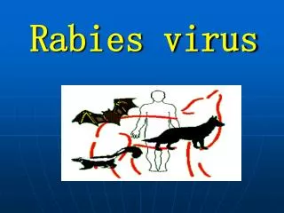
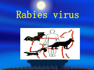
Rabies Transmission – by the bite of arabid animal (commonly a dog or a cat) – the contaminationof scratch woundsby virus- infectedsaliva. Clinical findings • Incubation/prodromal period 1 to 3 months • CNS infection • Hydrophobia. This fear of water is the result of the pain associated with drinking. • Seizures and hallucinations. • Paralysis may lead to respiratory failure. • Coma • Mortality is >99%. Most deadly virus!
Laboratory diagnosis Animals: Animals in a rabies endemic area should beheld for 10 days forobservation. If the animal is sick, kill the animal and collect samples from hippocampus tissues and look for 1) Negri bodies 2) viral antigens by immunofluorescence Human: Collect samples of saliva, serum, spinal fluid, and skin biopsies and perform RT-PCR or immunofluorescence. 海马回
Postexposure rabies prophylaxis Long incubation period is the basis of postexposure prophylaxis Local wound care Cleanse wound with soap and water. Sterilize with ethanol or iodine. Combination of passive and active immunization: Passive immunization with rabies antiserum(HRIG,given the day before or on the day of active immunization) Active immunization with antirabies vaccine (HDCV, im given on days 0, 3, 7, 14, 28).
From BSE to Prion Prion: An infectious proteinaceous particle of nonnucleic acid composition
Bovine spongiform encephalopathy (BSE) mad-cow disease
Prion protein (PrP): from PrPC to PrPSc Same amino acid sequences but different structures PrPSc (scrapie prion protein) 43% β- sheet and 30% α-helix Insoluble Resistant to protease K Pathogenic PrPC (cellular prion protein) Mainly α-helix Soluble Sensitive to protease K Has normal functions, not pathogenic
Prions aggregate extracellularly within the central nervous system to form plaques known as amyloid, which disrupt the normal tissue structure Strong resistance to autoclave, UV, regular disinfectants. Standard autoclave (121oC, 20 min) does not inactivate it. Needs 134oC, 2h
Transmissible SpongiformEncephalopathies (Prion Diseases) • Human – Kuru disease – Creutzfeldt--Jacob disease (CJD) – variant Creutzfeldt--Jacob disease (vCJD) • Animals – Scrapie of sheep and goats – Bovine spongiform encephalopathy (BSE, mad cow) Progressive neurodegenerative disease
Diagnosis and Prevention • Immunoblotting assay (Western blot): Treat tissue with proteinase K to digest PrPC, and then analyze with Western blot for the presence of PrPSc • Immunohistochemstry: Also treat with proteinase K first • Genetic analysis: For inherited mutations in the PrP gene. • Prevention: quarantine of animals and complete disinfection of medical devices. No treatment.