Download
1 / 3
30 likes | 319 Views
AAPOS poster 2006. Lateral Orbitotomy in the Management of Challenging Exotropia Yahalom C (1), Mc Nab A (2), Ben Simon G (2), Kowal L (2). 1-Hadassah University Hospital, Jerusalem. Israel. 2-Royal Victorian Eye and Ear Hospital, Melbourne, Australia.

E N D
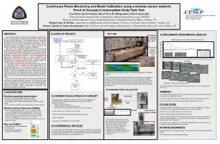
Lateral Orbitotomy in theManagement of Challenging ExotropiaYahalom C (1), Mc Nab A (2), Ben Simon G (2), Kowal L (2).1-Hadassah University Hospital, Jerusalem. Israel.2-Royal Victorian Eye and Ear Hospital, Melbourne, Australia • Introduction: The surgical management of recurrent exotropia (like the one seen following third nerve palsy and other cases with unwanted overactivity of the lateral rectus), that resists treatment by standard surgical techniques, is one of the most difficult problems facing the strabismus surgeon. The lateral rectus muscle often keeps pulling the eye back to exotropia following surgery, and further surgical procedures in this muscle become very hard. • Purpose: We present an “un-orthodox” surgical approach, to reach the posterior segment of the lateral rectus through a lateral orbitotomy, to manage these difficult cases of recurrent exotropia. • Methods: A review of the records of two patients with recurrent exotropia, following standard surgery was done. • Patient number one (N1) had an exotropia following retinal detachment repair, with failed multiple re-operations/explorations on his right lateral rectus (LR) which was super-glued to the globe due to extensive scarring (not feasible dissection from sclera). A lateral orbitotomy 1 was performed with lengthening of the posterior segment of the LR with a temporal fascia spacer. • Patient number 2 (N2) had a recurrent exotropia after a complete third nerve palsy. He underwent several surgeries to weaken the LR, including a failed trial to suturing LR to the orbital wall 2,3,4(due to shortened muscle after repeated surgeries), and an excision of anterior 10mm of the muscle 5 with myochol injection to the remaining posterior muscle. Two months following the surgery the eye was again 50 PD exotropic. An MRI showed reattachment of lateral rectus stump to the sclera near the level of the equator. Finally, we performed a posterior excision of LR remnants via a lateral orbitotomy. • Lateral Orbitotomy technique: A skin incision is made in the lateral canthal area, soft tissue is spread out down to the periosteum of the lateral orbital wall. A vertical incision is made in the lateral orbital rim periosteum, with peeling of this layer. The bone is cut using a saw superior to the level of the zygomatic arch and take off at the level of Whitnall's tubercle. The lateral wall is removed and kept in moist gauze. The periorbita is incised at the level of the lateral rectus muscle, the LR is lifted on a squint hook. • At the end of surgery the periorbita is sutured, the bone is positioned back in place and sutured to the wall. The periosteal lining is re-sutured. • Results: Both of the patients achieved satisfactory ocular alignment following surgery. These results were stable for 4 months in patient N1, and 2 years in patient N2. No mayor complications occurred. Any residual XT?/??? • Conclusion: Lateral orbitotomy for posterior lengthening/extirpation of lateral rectus in resistant exotropia, when a standard anterior approach for surgery is not feasible after repeated surgery on LR, is a safe and effective surgical procedure for restoring ocular alignment in persistent exodeviation. References: 1-Arai H et al. Lateral approach to intraorbital lesions: anatomic and surgical considerations. Neurosurgery 1996;39(6):1157-1163. 2-Velez F, Thacker N, Britt M, Alcorn D, Foster R, Rosenbaum A. Rectus muscle orbital wall fixation: a reversible profound weakening procedure.J AAPOS. 2004 Oct;8(5):473-80 3-Morad Y, Kowal L, Scott A. Lateral rectus muscle disinsertion and reattachment to the lateral orbital wall. BJO 2005;89:983-985. 4-Salazar-Leon JA, Ramirez-Ortiz MA, Salas-Vargas M. The surgical correction of paralytic strabismus using fascia lata. J Pediatr Ophthalmol Strabismus 1998;35:27-32. 5-Sato M, Maeda M, Ohmura T, et al. Myectomy of lateral rectus muscle for third nerve palsy. Jpn J Ophthalmol 2000;44:555-558.
Lateral Orbitotomy in the Management of Challenging ExotropiaYahalom C (1), Mc Nab A (2), Ben Simon G (2), Kowal L (2).1-Hadassah University Hospital, Jerusalem. Israel.2-Royal Victorian Eye and Ear Hospital, Melbourne, Australia Fig.1: patient N1 (Pre-op) Purpose: We present an “un-orthodox” surgical approach, to reach the posterior segment of the lateral rectus through a lateral orbitotomy, to manage these difficult cases of recurrent exotropia. Fig.2: LR muscle exposure through lateral orbitotomy Methods: A review of the records of two patients with recurrent exotropia, following standard surgery was done. Patient number one (N1) had an exotropia following retinal detachment repair, with failed multiple re-operations/explorations on his right lateral rectus (LR) which was super-glued to the globe due to extensive scarring (not feasible dissection from sclera). A lateral orbitotomy 1 was performed with lengthening of the posterior segment of the LR with a temporal fascia spacer. Patient number 2 (N2) had a recurrent exotropia after a complete third nerve palsy. He underwent several surgeries to weaken the LR, including a failed trial to suturing LR to the orbital wall 2,3,4(due to shortened muscle after repeated surgeries), and an excision of anterior 10mm of the muscle 5 with myochol injection to the remaining posterior muscle. Two months following the surgery the eye was again 50 PD exotropic. An MRI showed reattachment of lateral rectus stump to the sclera near the level of the equator. Finally, we performed a posterior excision of LR remnants via a lateral orbitotomy. Lateral Orbitotomy technique: A skin incision is made in the lateral canthal area, soft tissue is spread out down to the periosteum of the lateral orbital wall. A vertical incision is made in the lateral orbital rim periosteum, with peeling of this layer. The bone is cut using a saw superior to the level of the zygomatic arch and take off at the level of Whitnall's tubercle. The lateral wall is removed and kept in moist gauze. The periorbita is incised at the level of the lateral rectus muscle, the LR is lifted on a squint hook. At the end of surgery the periorbita is sutured, the bone is positioned back in place and sutured to the wall. The periosteal lining is re-sutured. Conclusion: Lateral orbitotomy for posterior lengthening/extirpation of lateral rectus in resistant exotropia, when a standard anterior approach for surgery is not feasible after repeated surgery on LR, is a safe and effective surgical procedure for restoring ocular alignment in persistent exodeviation. References: 1-Arai H et al. Lateral approach to intraorbital lesions: anatomic and surgical considerations. Neurosurgery 1996;39(6):1157-1163. 2-Velez F, Thacker N, Britt M, Alcorn D, Foster R, Rosenbaum A. Rectus muscle orbital wall fixation: a reversible profound weakening procedure.J AAPOS. 2004 Oct;8(5):473-80 3-Morad Y, Kowal L, Scott A. Lateral rectus muscle disinsertion and reattachment to the lateral orbital wall. BJO 2005;89:983-985. 4-Salazar-Leon JA, Ramirez-Ortiz MA, Salas-Vargas M. The surgical correction of paralytic strabismus using fascia lata. J Pediatr Ophthalmol Strabismus 1998;35:27-32. 5-Sato M, Maeda M, Ohmura T, et al. Myectomy of lateral rectus muscle for third nerve palsy. Jpn J Ophthalmol 2000;44:555-558. Results: Both of the patients achieved satisfactory ocular alignment following surgery. These results were stable for 8 months in patient N1, and 2 years in patient N2. No mayor complications occurred. Patient N1has a small residual XT Fig.3: Patient N1 (post-op) Introduction: The surgical management of recurrent exotropia (like the one seen following third nerve palsy and other cases with unwanted overactivity of the lateral rectus), that resists treatment by standard surgical techniques, is one of the most difficult problems facing the strabismus surgeon. The lateral rectus muscle often keeps pulling the eye back to exotropia following surgery, and further surgical procedures in this muscle become very hard.