Download
1 / 26
260 likes | 397 Views
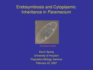
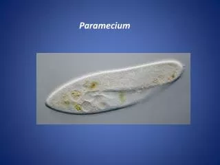
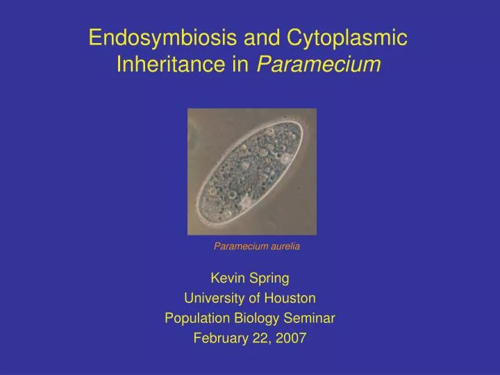
Endosymbiosis and Cytoplasmic Inheritance in Paramecium. Kevin Spring University of Houston Population Biology Seminar February 22, 2007. Paramecium aurelia. This presentation will focus on the following:. Altenburg paper (1948) Plasmagene hypothesis Kappa body symbiosis.

E N D
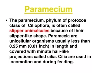
Endosymbiosis and Cytoplasmic Inheritance in Paramecium Kevin Spring University of Houston Population Biology Seminar February 22, 2007 Paramecium aurelia
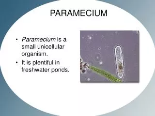
This presentation will focus on the following: • Altenburg paper (1948) • Plasmagene hypothesis • Kappa body symbiosis Current understanding of Kappa bodies (Preer 1974) Other cytoplasmic inheritance in Paramecium (Meyer 2002) • Paramecium biology • Cell biology • Life cycle
Altenburg paper (1948) investigates the evidence that Kappa bodies are a symbiont Kappa bodies are elements within Paramecium that cause themto be killers Killer Paramecium kill other Paramecium in the immediate environment Kappa particles, thought to be plasmagenes by Sonneborn, but Altenburg suggest they may be symbionts
The plasmagene theory suggested kappa bodies were genes within the cytoplasm Plasmagenes defined as self-replicating structure capable of producing traits that exist in the cytoplasm and are independent of chromosomal genes. The trait that Kappa bodies produce is the killing factor Kappa bodies are inherited through the cytoplasm and not through chromosomes Sonneborn wrote in 1976, “It was awful of me to be so attached to a pet idea. That was an ordeal between my mind and my heart and it took a while for the mind to win and the heart to accept. Impersonal scientific objectivity is a goal to be sought by hard self-discipline; we are not born with it.”
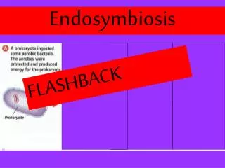
Altenburg’s evidence that Kappa bodies are symbionts is strongly supported by evidence Preer (1948) showed Kappa is large enough to see under a light microscope 38o C kills Kappa but not Paramecium Division of Kappa and Paramecium is independent of each other Paramecium with symbiont (2) There is an upper limit of the # of Kappa in Paramecium More likely a symbiont than a parasite
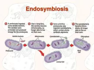
Preer (1974) reviewed the overwhelming evidence that Kappa bodies are symbionts Kappa contains DNA, RNA, protein, and lipids in proportions expected in bacteria Kappa contains electron transport system with cytochromes similar to bacteria and not eukaryotes Electron micrograph of symbionts (2) Electron microscopy clearly showed that Kappa is prokaryotic Electron micrograph of flagellated Kappa (2)
Current information has shown why Kappa induces killing and the different types of bacteria symbiosis Kappa bodies kill other Paramecium by releasing toxins into the environment The presence of the symbiont makes the host resistant to the toxin Kappa bodies are transmitted by the cytoplasm during asexual division gamma sigma lambda alpha Many other types of symbionts found delta pi Kappa is the most common omega mu
The discovery of bacterial symbionts within Paramecium allows for their taxonomic classification Kappa, mu, gamma, and nu are in genera Caedobacter Alpha bodies are in the genera Cytophaga Lambda and sigma are in genera Lyticum Delta bodies are in genera Tectobacter
Differences have been found between Kappa bodies in the same host Some Kappa bodies contain refractile ( R ) bodies R body is a type of inclusion body When genes from one organism are within another organism and are transcribed, a inactive protein may form Magnified image of coiled R body (2)
Kappa bodies may contain R bodies and it affects their reproductive capability Nonbright Kappa bodies do not contain R bodies but can reproduce Bright Kappa bodies do contain R bodies but cannot reproduce Dividing symbiont (2) Nonbrights produce other nonbrights, but occasionally a nonbright turns into a bright Toxicity associated only with Brights
There is still unsolved questions regarding Kappa body symbiosis What benefit does Paramecium get from the symbiosis? How does the presence of a Kappa body induce resistance to the toxin? Resistance can be overcome with large toxin dose The presence of Kappa with or without R bodies induces resistance to the toxin
Other types of cytoplasmic inheritance discovered in Paramecium and other ciliates is: Genome-wide DNA rearrangements Mating type Serotypes
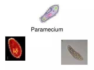
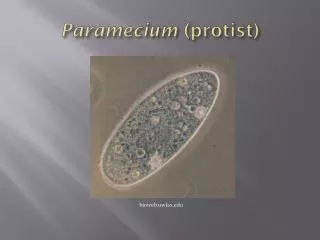
Paramecium has a complex cellular biology Eukaryotic Ciliates contain at least 2 nuclei Germ-line micronucleus (MIC) Somatic macronucleus (MAC) MAC is generated from the MIC Extensive genome rearrangements occur in the MAC Diagram of Paramecium (3)
The two nuclei make the life cycle of Paramecium more complicated than other eukaryotes MIC goes through meiosis and the haploid MIC goes through mitosis Result is 4 haploid MIC, but 2 are degraded Paramecium exchange 1 haploid MIC MIC fuse and form diploid MIC and duplicate via mitosis Old MAC degrades and duplicated MIC is processed into new MAC In asexual reproduction, the MIC goes through mitosis and the MAC goes through amitosis
Genome-wide rearrangements of the MAC genome consists of deletion of DNA sequences and chromosome amplification The developing new MAC loses 10 - 95% of the genome depending on the ciliate MAC chromosomes are amplified to a high ploidy level Deletion occurs after an initial amplification of the MIC genome but before the ploidy level is reached
The deletion of DNA is located at specific sequences called internal excised sequences (IES) IES are located in coding and noncoding regions of the MIC genome These sequences are not present in the MAC genome At some point in MAC development, the IES sequences are deleted How is IES deletion maternally inherited?
The mating type of Paramecium shows maternal inheritance Conjugation of P. caudatum by Yanagi Paramecium has 2 mating types - O and E Both are not determined by genetic differences as they are both produced in homozygous wild-type strains Mating type is the same through asexual reproduction but can change after sexual conjugation and MAC formation After conjugation O cells mostly produce other O cells and E cells produce other E cells
Paramecium mating types do not follow the Mendelian segregation of alleles Mendelian segregation of allelic pairs Maternal inheritance of mating types (4)
Mating types O and E depends on different states of MAC genome Transferring E maternal MAC into O cell causes the progeny to become E Transferring O MAC does not change E cells O is the default mating type O cell E cell E cell Produces Insert E MAC
This differential state of MAC is dependent on the presence of IES in the MAC The mutation mTFEcauses O cells to become E This mutation affects the excision of an IES on the G gene The G gene is a surface antigen and the failure of excision causes a nonfunctional protein to be translated Functional - type O excision Mutational retention Nonfunctional - type E MIC G gene MAC G gene
Microinjection studies have shown that the presence of an IES sequence in the MAC inhibits the excision of its homologous IES in the MIC O cells contain G gene in the MAC without its IES (IES-) E cells contain the G gene in the MAC with its IES (IES+) Injecting a plasmid of IES+ G gene into O cell’s MAC created the retention of the IES in the MAC of daughter cells Injection of IES- plasmid did not induce excision The presence of IES in the MAC causes the retention of the IES in subsequent generations after sexual conjugation
Microinjection of IES+ plasmid retains the IES in the MAC genome after autogamy
Meyer (2002) asked, “How can a sequence introduced in one nucleus affect the excision of the homologous sequence in another nucleus?” Two models developed Model 1: Sequence-specific protein factors are required for the excision of the IES in the developing MAC The problem with this model is the large number of protein factors needed, about 50,000 Model 2: Sequence specificity is achieved by homologous nucleic acid (most likely RNA) that is transported from the maternal MAC to the developing MAC
Mochizuki (2004) explained the Scanning Model, a synthesis of Meyer’s model 1 and 2 Entire MIC genome is transcribed bi-directionally and forms dsRNA dsRNA is cut up into smaller RNA called scnRNA scnRNA move to the old MAC and any matching homologous sequences are degraded scnRNA that were not degraded move to the developing MAC These scnRNAs target homologous sequences which are deleted in an RNAi-like mechanism
Summary Paramecium has many instances of cytoplasmic and maternal inheritance Paramecium (6) Kappa bodies are bacterial symbionts that produce a killing factor and they are inherited through the cytoplasm Electron micrograph of Kappa (2) IES excision and retention in the MAC is maternally inherited by the genome present in the MAC
References 1.Altenburg E (1948) The role of symbionts and autocatalysts in the genetics of the ciliate. The American Naturalist, 82: 252-264. 2.Preer JR, Preer LB and Jurand A (1974) Kappa and other endosymbionts in Paramecium aurelia. Bacteriological Reviews, 38: 113-163. 3.Spark Notes. Protist. http://www.sparknotes.com/biology/microorganisms/protista/section2.rhtml. 4.Meyer E and Garnier O (2002) Non-Mendelian inheritance and homology-dependent effects in ciliates. Advances in Genetics, 46: 305-337. 5.Mochizuki K and Gorovsky MA (2004) Small RNAs in genome rearrangements in Tetrahymena. Current Opinions in Genetics and Development, 14: 181-187. 6.Ken Todar’s Microbial World. Introduction to the Microbial World. http://www.bact.wisc.edu/themicrobialworld/paramecium.jpg. 7. 7. Preer JR (2006) Perspectives: anecdotal, historical and critical commentaries on genetics. Genetics, 172: 1373-1377