Download

1 / 87
1.51k likes | 3.64k Views
LYMPHATIC DRAINAGE OF HEAD & NECK. CONTENTS. Introduction History of lymphatic system Development of lymphatic system Lymph Lymph node Lymph nodes of head and neck Examination on neck nodes Cervical lymphadenopathy Refrences. INTRODUCTION.

E N D
CONTENTS • Introduction • History of lymphatic system • Development of lymphatic system • Lymph • Lymph node • Lymph nodes of head and neck • Examination on neck nodes • Cervical lymphadenopathy • Refrences
INTRODUCTION • Lymphatic system consist of fluid called LYMPH • DEFINITION:Transparent, slightly yellowish liquid of alkaline reaction found in lymphatic vessel and derived from tissue fluid • Lymphatic system is absent in: -C.N.S. -Cornea -Superficial layer of skin -bones -alveoli of lung
HISTORY OF LYMPH DISCOVERY AND LYMPHATIC DRAINAGE • In 1650,John Paquet-cysternachyli • In 1962,Gaspard Asseli -milky veins • OlaufRudbeck-first person to describe the lymphatic system • Alexander of winiwater-protocol for draining lymphedenomas • F.D.Millard -diagnostic importance by palpating lymphatic gland • Emil Vodder -technoque of lymphatic dranaige • BronoChilky-rhythm of lymphatic flow
DEVELOPMENT • Starts at 5th week of intrauterine life. • First signs of lymphatic system are seen in the form of a number of endothelium lined lymph sacs
SIX PRIMARY LYMPH SACS ARE FORMED. 2 Jugular sacs (right and left) • At the junction of subclavian and anterior cardinal veins. 2 iliac sac (right and left) • At the junction of the iliac and posterior cardinal vein. Retroperitonial sac (Unpaired) • Near the root of the mesentery. Cisterna chyli (unpaired) • Dorsal to retroperitonial sac All the sacs except the cisternachyli are invaded by connective tissue and lymphocytes and are converted into lymph nodes
Rate of lymph flow: About 120ml of lymph flows into blood
Rate of flow of lymph along the human thoracic duct is from 1-1.5ml/min. • Regulation of the lymph flow mainly depends upon : • Interstitial pressure • Atrial pulsation • Intrathorasic pressure • Muscular massage
FORMATION OF LYMPH • Lymph is formed from tissue fluid,anything that increases amount of tissue fluid, will increase the rate of lymph formation • Various mechanisms: • Filteration from plasma normally exceeds resorption leading to net formation of tissue fluid • Increase in interstitial fluid hydrostatic pressure favouring the movement of tissue fluid into lymphatic capillary forming lymph
FUNCTIONS OF THE LYMPH • Nutritive • Drainage • Transmission of proteins • Absorption of fats • Defensive
LYMPH MOVEMENT • It takes place with the help of: • Contractile skeletal muscle • Presence of valve • Contraction of smooth muscle in large lymphatic trunk • Pressure change in muscle during breathing
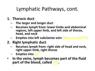
LYMPHATIC PATHWAYS • FLOW CHART LYMPHATIC CAPPILLARY LYMPHATIC VESSEL LYMPHATIC NODE LYMPHATIC VESSEL LYMPHATIC TRUNK SUBCLAVIAN VEIN
Before Lymph is returned to the blood stream, it passes through at least one lymph node and often through several • The Lymph vessels that carry lymph to a lymph node are referred to as afferent & those that transport it away from a node are called efferent vessels
STRUCTURE OF LYMPH NODE • Lymph nodes are oval-shaped of bean-shaped structures • Some are as small as a pinhead and others as large as a lima bean • Each lymph node is enclosed by a fibrous capsule • Once lymph enters the node, it "percolates" slowly through the spaces known as sinuses before draining into a single efferent draining vessel. • One-way valves in both the afferent and efferent vessels keep lymph flowing in one direction
Fibrous septa or trabeculae extend from the covering capsule toward the center of the node. • Cortical nodules found within the sinuses along the outer region of the node are separated from each other by these trabeculae. • Each cortical nodule is composed of packed lymphocytes that surround a less dense area called a germinal center. • When an infection is present, germinal centers form and the node begins to release lymphocytes.
Lymphocytes begin their final stages of maturation within the germinal center of the nodule and then are pushed to the more densely packed outer layers as they mature to become antibody-producing plasma cells. • The center or medulla of a lymph node is composed of sinuses and cords. • Both the cortical and medullary sinuses are lined with specialized reticuloendothelial cells (fixed macrophages) which are capable of phagocytosis
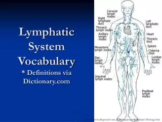
CLASSIFICATION Upper horizontal chain of nodes: • Submental • Submandibular • Parotid • Postauricular • Occipital
SUBMENTAL NODES • Lie on mylohyoid muscle in the submental triangle • 2 to 8 in number • Drainage –afferents come from the chin, middle part of lower lip, anterior gums, anterior floor of mouth and tip of tongue. • Efferents -they go to submandibular and internal jugular chain
SUBMANDIBULAR NODES • They lie in submandibular triangle in relation to submandibular gland. • Afferents come from lateral part of the lower lip, upper lip, cheek,nasal vestibule and anterior part of nasal cavity, gums,teeth medial canthus, soft palate, anterior pillar, anterior part of tongue, submandibular and sublingual salivary glands and floor of mouth • Efferents go to internal jugular chain
PAROTID NODES • They lie in relation to the parotid salivary gland. • Afferents come from the scalp,pinna, external auditory canal,face,buccal mucosa. • Efferents go to internal jugular or external jugular chain
POST AURICULAR NODES • Also called as mastoid nodes • They lie behind the thepinna over the mastoid. • Afferents come from the scalp, posterior surface of pinna and skin of mastoid. • Efferents drain into internal jugular chain
OCCIPITAL NODES • They lie at the apex of the posterior triangle • Afferents come from scalp, skin of upper neck. • Efferents drain into upper accessory chain of nodes
Lateral cervical nodes • They include nodes, superficial and deep to sternocleidomastoid muscle and in the posterior triangle. • Superficial external jugular group • Deep group i. Internal jugular chain (upper,middle and lower groups) ii. Spinal accessory chain iii. Transverse cervical chain
LATERAL CERVICAL NODES • a) Superficial group – it lies along external jugular vein and drains into internal jugular and transverse cervical nodes • b)Deep group It consists of three chains, the internal jugular, spinal accessory and transverse cervical
Internal jugular chain • Lymph nodes of internal jugular chain lie anterior, lateral and posterior to internal jugular vein. • Upper group (jugulodigastric node) – drains oral cavity, orpharynx, nasopharynx,hypopharynx, larynx and parotid. • Middle group drains hypopharynx, larynx, throid, oral cavity, oropharynx. • Lower jugular group drains larynx, thyroid and cervical oesophagus
Spinal accessory chain • Lies along the spinal accessory nerve. Spinal accessory chain drains the scalp, skin of the neck, the nasopharynx, occipital and postauricular nodes. • Efferents from this chain drain into transverse cervical chain
Transverse cervical chain (supraclavicular nodes) • It lies horizontally, along the trasverse cervical vessels, in thelower part of the posterior triangle. • The medial nodes of the group are called scalene nodes. • Afferents to those nodes come from the accessory chain and also infraclavicularstructures,e.g. breast, lung, stomach, colon, ovary and testis
Anterior cervical nodes • Anterior jugular chain • Juxtavisceral chain i. Prelaryngeal ii. Pretracheal iii. Paratracheal
ANTERIOR CERVICAL NODES • They lie between the two carotids and below the level of hyoid bone and consist of two chains: (a) Anterior jugular chian • It lies along anterior jugular vein and drains the skin of anterior neck. (b) Juxtavisceral chain • It consists of prelaryngeal,pretracheal and paratracheal nodes • Prelaryngeal node (Delphian node)-lies on cricothyroid membrane and drains subgottic region of larynx and pyriform sinuses • Pretracheal nodes lie in front of the trachea, and drain thyroid gland and the trachea.Efferents from these nodes go to paratracheal, lower internal jugular and anterior mediastinal nodes • Paratracheal Nodes – drain the thyroid lobes, subglottic larynx, tracha and cervical oesophagus
CLASSIFICATION OF NECK NODES ACCORDING TO LEVELS • Level I Submental (IA) Submandibular (IB) • Level II Upper jugular • Level III middle jugular • Level IV Lower jugular • Level V Posterior triangle group(Spinal accessory and transverse cervical chains) • Level VI Prelaryngeal Pretracheal Paratracheal • Level VII Nodes of upper mediastinum
Level I includes : • IA Submental nodes, which lie in the submental triangle i.e. between right and left anterior bellies of diagastric muscles and the hyoid bone. • IB Submandibular ones, lying between anterior and posterior bellies of diagastric muscle and the body of mandible