Download
1 / 44
440 likes | 541 Views
Cells and Tissues. Standard- SAP1. Students will analyze anatomical structures in relationship to their physiological functions. d. Relate cellular metabolism and transport to homeostasis and cellular reproduction

E N D
Standard- SAP1. Students will analyze anatomical structures in relationship to their physiological functions. • d. Relate cellular metabolism and transport to homeostasis and cellular reproduction • e. Describe how structure and function are related in terms of cell and tissue types.
Questions • Essential Question(s): How are anatomical structures (body parts) related to physiological Functions (how they move)? • Key Questions: • What are organelles? • What are the different organelles and their functions? • What are the different types of cells? • What are the tissue types? • What is cellular metabolism? • What is cellular transport? • How do cellular metabolism and cellular transport affect homeostasis & cellular reproduction?
Cell Theory • All cells come from pre-existing cells • Cell are the basic unit of structure and function • All living things are composed of cells
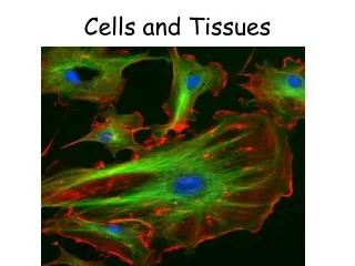
Cells and Tissues • Carry out all chemical activities needed to sustain life • Cells are the building blocks of all living things • Tissues are groups of cells that are similar in structure and function
Anatomy of the Cell • Cells are not all the same • All cells share general structures • All cells have three main regions • Nucleus • Cytoplasm • Plasma membrane Figure 3.1a
The Nucleus • Control center of the cell • Contains genetic material (DNA) • Three regions • Nuclear envelope (membrane) • Nucleolus • Chromatin
The Nucleus Figure 3.1b
The Nucleus • Nuclear envelope (membrane) • Barrier of the nucleus • Consists of a double membrane • Contains nuclear pores that allow for exchange of material with the rest of the cell
The Nucleus • Nucleoli • Nucleus contains one or more nucleoli • Sites of ribosome assembly • Ribosomes migrate into the cytoplasm through nuclear pores
The Nucleus • Chromatin • Composed of DNA and protein • Present when the cell is not dividing • Scattered throughout the nucleus • Condenses to form chromosomes when the cell divides
Plasma Membrane • Barrier for cell contents • Double phospholipid layer • Hydrophilic heads • Hydrophobic tails • Also contains proteins, cholesterol, and glycoproteins
Plasma Membrane PLAY Membrane Structure Figure 3.2
Plasma Membrane Specializations • Microvilli • Finger-like projections that increase surface area for absorption
Plasma Membrane Specializations • Membrane junctions • Tight junctions • Impermeable junctions • Bind cells together into leakproof sheets • Desmosomes • Anchoring junctions that prevent cells from being pulled apart • Gap junctions • Allow communication between cells
Plasma Membrane Specializations Figure 3.3
Plasma Membrane Specializations PLAY Tight Junctions PLAY Desmosomes (Anchoring Junctions)
Cytoplasm • Cytoplasm is the material outside the nucleus and inside the plasma membrane
Cytoplasm • Contains three major elements • Cytosol • Fluid that suspends other elements • Organelles • Metabolic machinery of the cell • “Little organs” that perform functions for the cell • Inclusions • Chemical substances such as stored nutrients or cell products
Cytoplasmic Organelles Figure 3.4
Cytoplasmic Organelles • Mitochondria • “Powerhouses” of the cell • Change shape continuously • Carry out reactions where oxygen is used to break down food • Provides ATP for cellular energy
Cytoplasmic Organelles • Ribosomes • Made of protein and RNA • Sites of protein synthesis • Found at two locations • Free in the cytoplasm • As part of the rough endoplasmic reticulum
Cytoplasmic Organelles • Endoplasmic reticulum (ER) • Fluid-filled tubules for carrying substances • Two types of ER • Rough endoplasmic reticulum • Studded with ribosomes • Synthesizes proteins • Smooth endoplasmic reticulum • Functions in lipid metabolism and detoxification of drugs and pesticides
As the protein is synthesizedon the ribosome, it migratesinto the rough ER cistern. Ribosome mRNA Rough ER In the cistern, the protein foldsinto its functional shape. Shortsugar chains may be attachedto the protein (forming aglycoprotein). Protein The protein is packaged in atiny membranous sac called atransport vesicle. Transportvesicle buds off The transport vesicle buds fromthe rough ER and travels to theGolgi apparatus for furtherprocessing or goes directly tothe plasma membrane where itscontents are secreted. Protein insidetransport vesicle Rough Endoplasmic Reticulum Figure 3.5
As the protein is synthesizedon the ribosome, it migratesinto the rough ER cistern. Ribosome mRNA Rough ER Protein Rough Endoplasmic Reticulum Figure 3.5, step 1
As the protein is synthesizedon the ribosome, it migratesinto the rough ER cistern. Ribosome mRNA Rough ER In the cistern, the protein foldsinto its functional shape. Shortsugar chains may be attachedto the protein (forming aglycoprotein). Protein Rough Endoplasmic Reticulum Figure 3.5, step 2
As the protein is synthesizedon the ribosome, it migratesinto the rough ER cistern. Ribosome mRNA Rough ER In the cistern, the protein foldsinto its functional shape. Shortsugar chains may be attachedto the protein (forming aglycoprotein). Protein The protein is packaged in atiny membranous sac called atransport vesicle. Transportvesicle buds off Rough Endoplasmic Reticulum Figure 3.5, step 3
As the protein is synthesizedon the ribosome, it migratesinto the rough ER cistern. Ribosome mRNA Rough ER In the cistern, the protein foldsinto its functional shape. Shortsugar chains may be attachedto the protein (forming aglycoprotein). Protein The protein is packaged in atiny membranous sac called atransport vesicle. Transportvesicle buds off The transport vesicle buds fromthe rough ER and travels to theGolgi apparatus for furtherprocessing or goes directly tothe plasma membrane where itscontents are secreted. Protein insidetransport vesicle Rough Endoplasmic Reticulum Figure 3.5, step 4
Cytoplasmic Organelles • Golgi apparatus • Modifies and packages proteins • Produces different types of packages • Secretory vesicles • Cell membrane components • Lysosomes
Cytoplasmic Organelles • Lysosomes • Contain enzymes that digest worn-out or nonusable materials within the cell
Cytoplasmic Organelles • Peroxisomes • Membranous sacs of oxidase enzymes • Detoxify harmful substances such as alcohol and formaldehyde • Break down free radicals (highly reactive chemicals) • Replicate by pinching in half
Cytoplasmic Organelles • Cytoskeleton • Network of protein structures that extend throughout the cytoplasm • Provides the cell with an internal framework Figure 3.7a
Cytoplasmic Organelles • Cytoskeleton • Three different types of elements • Microfilaments (largest) • Intermediate filaments • Microtubules (smallest) Figure 3.7b–d
Cytoplasmic Organelles • Centrioles • Rod-shaped bodies made of microtubules • Direct the formation of mitotic spindle during cell division
Cellular Projections • Not found in all cells • Used for movement • Cilia move materials across the cell surface • Located in the respiratory system to move mucus • Flagella propel the cell • The only flagellated cell in the human body is sperm
STOP • Complete Cell Organelle Worksheet
Cell Diversity Figure 3.8a
Cell Diversity Figure 3.8b
Cell Diversity Figure 3.8c
Cell Diversity Figure 3.8d
Cell Diversity Figure 3.8e
Cell Diversity Figure 3.8f
Cell Diversity Figure 3.8g
A Tour of the Cell PLAY Tour of the Cell