Download
1 / 24
380 likes | 1.5k Views
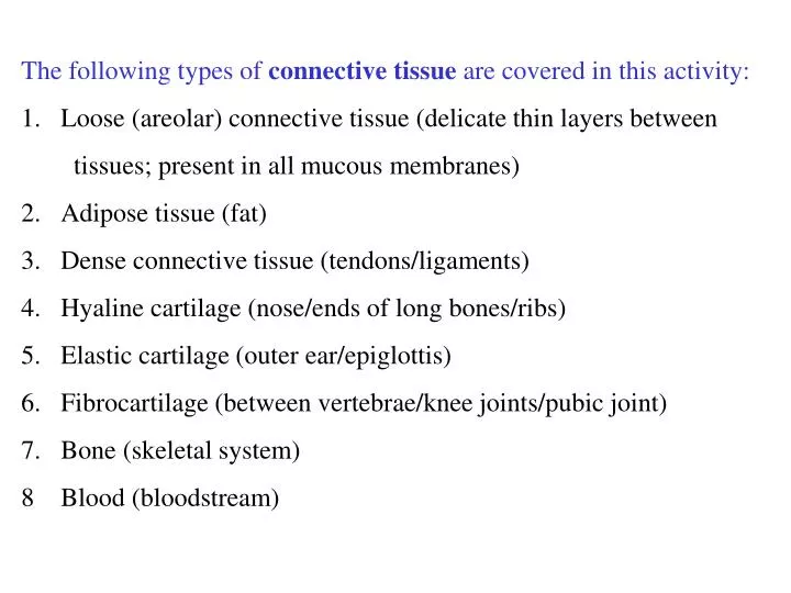
The following types of connective tissue are covered in this activity: Loose (areolar) connective tissue (delicate thin layers between tissues; present in all mucous membranes) 2. Adipose tissue (fat) 3. Dense connective tissue (tendons/ligaments)

E N D
The following types of connective tissue are covered in this activity: • Loose (areolar) connective tissue (delicate thin layers between • tissues; present in all mucous membranes) • 2. Adipose tissue (fat) • 3. Dense connective tissue (tendons/ligaments) • 4. Hyaline cartilage (nose/ends of long bones/ribs) • 5. Elastic cartilage (outer ear/epiglottis) • 6. Fibrocartilage (between vertebrae/knee joints/pubic joint) • 7. Bone (skeletal system) • 8 Blood (bloodstream)
Table 4.1 Comparison of Classes of Connective Tissues (1 of 2)
Table 4.1 Comparison of Classes of Connective Tissues (2 of 2)
Figure 4.8a Connective tissues. (a) Connective tissue proper: loose connective tissue, areolar Description:Gel-like matrix with all three fiber types; cells: fibroblasts, macrophages, mast cells, and some white blood cells. Elastic fibers Function: Wraps and cushions organs; its macrophages phagocytize bacteria; plays important role in inflammation; holds and conveys tissue fluid. Collagen fibers Location: Widely distributed under epithelia of body, e.g., forms lamina propria of mucous membranes; packages organs; surrounds capillaries. Fibroblast nuclei Epithelium Photomicrograph: Areolar connective tissue, a soft packaging tissue of the body (300x). Lamina propria
Figure 4.7 Areolar connective tissue: A prototype (model) connective tissue. Cell types Extracellular matrix Ground substance Fibers • Collagen fiber • Elastic fiber • Reticular fiber Macrophage Fibroblast Lymphocyte Fat cell Capillary Mast cell Neutrophil
Figure 4.8b Connective tissues. (b) Connective tissue proper: loose connective tissue, adipose Description: Matrix as in areolar, but very sparse; closely packed adipocytes, or fat cells, have nucleus pushed to the side by large fat droplet. Function: Provides reserve food fuel; insulates against heat loss; supports and protects organs. Nucleus of fat cell Location: Under skin in the hypodermis; around kidneys and eyeballs; within abdomen; in breasts. Vacuole containing fat droplet Adipose tissue Photomicrograph: Adipose tissue from the subcutaneous layer under the skin (350x). Mammary glands
Figure 4.8c Connective tissues. (c) Connective tissue proper: loose connective tissue, reticular Description: Network of reticular fibers in a typical loose ground substance; reticular cells lie on the network. Function: Fibers form a soft internal skeleton (stroma) that supports other cell types including white blood cells, mast cells, and macrophages. White blood cell (lymphocyte) Location: Lymphoid organs (lymph nodes, bone marrow, and spleen). Reticular fibers Spleen Photomicrograph: Dark-staining network of reticular connective tissue fibers forming the internal skeleton of the spleen (350x).
Figure 4.8d Connective tissues. (d) Connective tissue proper: dense connective tissue, dense regular Description: Primarily parallel collagen fibers; a few elastic fibers; major cell type is the fibroblast. Collagen fibers Function: Attaches muscles to bones or to muscles; attaches bones to bones; withstands great tensile stress when pulling force is applied in one direction. Location: Tendons, most ligaments, aponeuroses. Nuclei of fibroblasts Shoulder joint Ligament Photomicrograph: Dense regular connective tissue from a tendon (500x). Tendon
Figure 4.8e Connective tissues. (e) Connective tissue proper: dense connective tissue, dense irregular Description: Primarily irregularly arranged collagen fibers; some elastic fibers; major cell type is the fibroblast. Nuclei of fibroblasts Function: Able to withstand tension exerted in many directions; provides structural strength. Location: Fibrous capsules of organs and of joints; dermis of the skin; submucosa of digestive tract. Collagen fibers Fibrous joint capsule Photomicrograph: Dense irregular connective tissue from the dermis of the skin (400x).
Figure 4.8f Connective tissues. (f) Connective tissue proper: dense connective tissue, elastic Description: Dense regular connective tissue containing a high proportion of elastic fibers. Function: Allows recoil of tissue following stretching; maintains pulsatile flow of blood through arteries; aids passive recoil of lungs following inspiration. Elastic fibers Location: Walls of large arteries; within certain ligaments associated with the vertebral column; within the walls of the bronchial tubes. Aorta Photomicrograph: Elastic connective tissue in the wall of the aorta (250x). Heart
Figure 4.8g Connective tissues. (g) Cartilage: hyaline Description: Amorphous but firm matrix; collagen fibers form an imperceptible network; chondroblasts produce the matrix and when mature (chondrocytes) lie in lacunae. Function: Supports and reinforces; has resilient cushioning properties; resists compressive stress. Location: Forms most of the embryonic skeleton; covers the ends of long bones in joint cavities; forms costal cartilages of the ribs; cartilages of the nose, trachea, and larynx. Chondrocyte in lacuna Matrix Costal cartilages Photomicrograph: Hyaline cartilage from the trachea (750x).
Figure 4.8h Connective tissues. (h) Cartilage: elastic Description: Similar to hyaline cartilage, but more elastic fibers in matrix. Function: Maintains the shape of a structure while allowing great flexibility. Chondrocyte in lacuna Location: Supports the external ear (pinna); epiglottis. Matrix Photomicrograph: Elastic cartilage from the human ear pinna; forms the flexible skeleton of the ear (800x).
Figure 4.8i Connective tissues. (i) Cartilage: fibrocartilage Description: Matrix similar to but less firm than that in hyaline cartilage; thick collagen fibers predominate. Function: Tensile strength with the ability to absorb compressive shock. Location: Intervertebral discs; pubic symphysis; discs of knee joint. Chondrocytes in lacunae Intervertebral discs Collagen fiber Photomicrograph: Fibrocartilage of an intervertebral disc (125x). Special staining produced the blue color seen.
Figure 4.8j Connective tissues. (j) Others: bone (osseous tissue) Description: Hard, calcified matrix containing many collagen fibers; osteocytes lie in lacunae. Very well vascularized. Central canal Function: Bone supports and protects (by enclosing); provides levers for the muscles to act on; stores calcium and other minerals and fat; marrow inside bones is the site for blood cell formation (hematopoiesis). Lacunae Lamella Location: Bones Photomicrograph: Cross-sectional view of bone (125x).
Figure 4.8k Connective tissues. (k) Others: blood Description: Red and white blood cells in a fluid matrix (plasma). Plasma Function: Transport of respiratory gases, nutrients, wastes, and other substances. Neutrophil Location: Contained within blood vessels. Red blood cells Lymphocyte Photomicrograph: Smear of human blood (1860x); two white blood cells (neutrophil in upper left and lymphocyte in lower right) are seen surrounded by red blood cells.
Given the previous outline and examples Can you name? First, the tissue type Second, where in the body the tissue is found
What kind of tissue does this represent? Loose (areolar) connective tissue Where in the body can you find this tissue? delicate thin layers between tissues; present in all mucous membranes
What kind of tissue does this represent? Adipose tissue Where in the body can you find this tissue? fat
What kind of tissue does this represent? Dense connective tissue Where in the body can you find this tissue? tendons; ligaments
What kind of tissue does this represent? Hyaline cartilage Where in the body can you find this tissue? nose; ends of long bones; ribs
What kind of tissue does this represent? Elastic cartilage Where in the body can you find this tissue? outer ear; epiglottis
What kind of tissue does this represent? Fibrocartilage Where in the body can you find this tissue? between vertebrae; knee joints; pubic joint
What kind of tissue does this represent? Bone Where in the body can you find this tissue? skeletal system
What kind of tissue does this represent? Blood Where in the body can you find this tissue? bloodstream