Download
1 / 46
520 likes | 707 Views
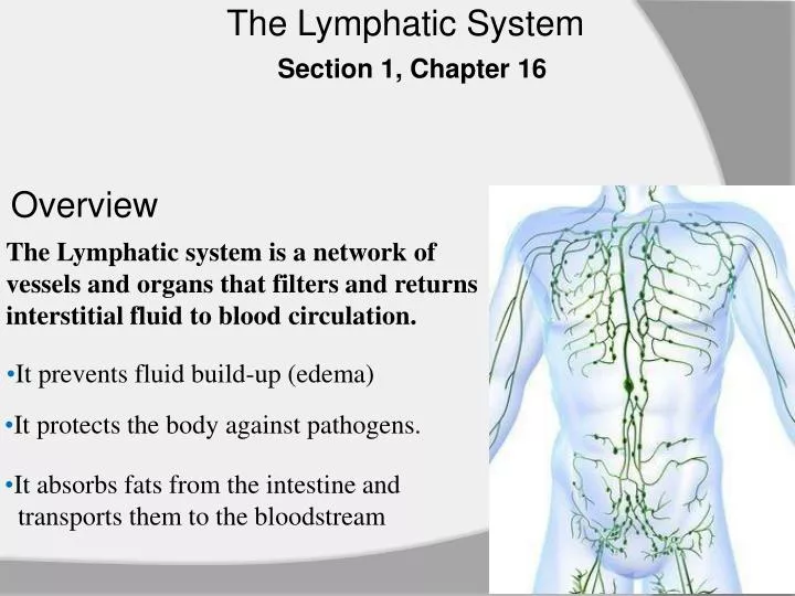
The Lymphatic System. Section 1, Chapter 16. Overview. The Lymphatic system is a network of vessels and organs that filters and returns interstitial fluid to blood circulation. . It prevents fluid build-up (edema). It protects the body against pathogens. .

E N D
The Lymphatic System Section 1, Chapter 16 Overview The Lymphatic system is a network of vessels and organs that filters and returns interstitial fluid to blood circulation. • It prevents fluid build-up (edema) • It protects the body against pathogens. • It absorbs fats from the intestine and • transports them to the bloodstream
Lymphatic Pathways • The lymphatic system begins at lymphatic capillaries • Lymphatic capillaries convey lymph towards • lymphatic vessels • Lymph passes through lymph nodes, where it • is filtered and monitored for pathogens. • Large lymphatic vessels (collecting ducts) ultimately return lymph to venous circulation.
Lymphatic Capillaries • Lymphatic capillaries are closed-ended vessels closely interlaced with blood capillaries. • They collect fluid forced out of blood capillaries (interstitial fluid) • and return it to venous circulation.
Lymphatic Capillaries Structure • Simple squamous epithelium (endothelium) anchored to connective tissue.
Formation of Lymph Osmotic pressure Hydrostatic pressure Hydrostatic pressure moves plasma out of blood capillaries into the interstitial space. Osmotic pressure only reabsorbs some of the lost fluid from interstitial space. Blood capillary To venule From arteriole l; Excess fluid enters through one-way valves lymph Interstitial space To Lymphatic vessel Lymphatic capillary
Lymphatic Vessels Structure: Lymphatic vessels are similar to veins, but thinner. 3 Layers: Tunica interna – endothelium and one-way valves Tunica media – smooth muscles and elastic fibers Tunica externa – connective tissue lymph Lymphatic Vessel with a flap-like valve. Arrow shows direction of lymph flow.
Lymphatic Trunks • Larger vessels lead to lymph nodes and then to larger lymphatic trunks • Lymphatic trunks are named for the body region they drain. • Lumbar Trunk – drains lymph from lower limbs and pelvic organs • Intestinal Trunk– drains abdominal viscera • Bronchomediastinal Trunk – drains portions of the thorax • Subclavian Trunk – drains upper limbs • Jugular Trunk – drains portions of the neck and head
Collecting Ducts Two Collecting ducts empty lymph from lymphatic trunks into venous circulation. • Thoracic Duct • Originates at the abdomen and empties into the left subclavian vein. • Drains lymph from entire lower body: lumbar and intestinal trunks • Also drains lymph from left upper limb, and left head and neck. • Right Lymphatic Duct • Originates in right thorax and empties into the right subclavian vein. • Drains lymph from right upper limb, and right head and neck.
Collecting Ducts Figure 16.6 Lymphatic pathways. The right lymphatic duct drains lymph from the upper right side of the body (in red), whereas the thoracic duct drains lymph from the rest of the body.
The Lymphatic Pathway Figure 16.7 The lymphatic pathway. This pathway is true for both right and left sides.
Lymph Movement • Muscle activity largely influences the movement of lymph through the lymphatic vessels via: • Action of skeletal muscles • Respiratory movements • Smooth muscle in the larger lymphatic vessels • Valves in the lymphatic vessels
Lymph Nodes • Lymph nodes are located along the lymphatic pathways • They contain lymphocytes and macrophages to fight invading pathogens • Lymph enters and leaves a lymph node through lymphatic vessels. • Afferent Vessels –convey lymph towards lymph node • Efferent Vessels – convey lymph away from lymph node.
Lymph Node Structure • Capsule of connective tissue (c.t.). • Trabiculae = c.t. partitions • Sinuses • Complex channels through which lymph circulates • Interlaced with reticular fibers. • Hilum • Entrance for blood vessels and nerves • Efferent vessels leave from the hilum Cortex (outer layer) • Nodules = dense populations of B-cells • and macrophages trabiculae Medulla (inner layer) • Reticulum • T-cells
Locations of Lymph Nodes There are about 450 lymph nodes throughout the body. • Cervical region – filter lymph from the face and scalp • Axillary region – filters lymph from upper limbs, thorax, and mammary glands • Supratrochlearregion – filter lymph from forearms • Enlarges in children in response to cuts on the hand • Inguinal region – filters lymph from lower limbs and external genitalia • Pelvic cavity – filter lymph from internal pelvic organs • Abdominal cavity –filter lymph from abdominal viscera • Thoracic cavity – filter lymph from thoracic viscera
Locations of Lymph Nodes Functions of Lymph Nodes Filter particles from lymph before returning it to the blood stream Monitor body fluids (immune surveillance) Lymphocyte production. Figure 16.11 Major locations of lymph nodes
Obstruction of Lymph Flow Axillary lymph nodes are often removed during a mastectomy to prevent the metastasis of breast cancer to other regions in the body. This can obstruct the drainage of lymph from the upper arm, resulting in edema.
MALT Mucosa-associated lymphoid tissue (MALT) – lymphatic nodules associated with the mucous membranes (i.e. digestive, respiratory, genitourinary tracts) Tonsils – encapsulated lymphoid tissues Peyer’s Patches – common in the ilium portion of the small intestine Appendix – contains aggregations of lymphatic nodules Individual lymphatic nodules are also found along the mucosal membrane. Two lymphatic nodules of the Peyer’s Patches in the small intestine.
Thymus • The thymus • Is enlarged in childhood, then atrophies in adult hood. • It is the site of T-cell production • Secretes Thymosin, which promotes the maturation of T-cells. Figure 16.13 Compared to other thoracic organs, the thymus in the fetus is large, but in the adult is small. Figure is not to scale.
The Spleen The spleen is the largest lymphatic organ. It is located in the upper left abdominal quadrant, just inferior to the diaphragm. The spleen’s primary function is to filter blood, remove old red blood cells, and house lymphocytes. Its structure resembles a large lymph node.
The Spleen • Capsule of connective tissue (c.t.). • Trabiculae = c.t. partitions • Hilum • Entrance for blood vessels and nerves • Venous Sinuses • Complex channels through which blood circulates • Interlaced with reticular fibers. • White Pulp • Lymphatic Nodules (packed with lymphocytes) • Red Pulp • Abundant Red Blood Cells (RBCs) • Removes old RBCs from circulation • Contains many lymphocytes and macrophages
ivyanatomy.com Section 2, Chapter 16 Lymphatic System & Defense Against Infection Pathogens that infiltrate the body may cause infection. • Pathogens Include: • Bacteria, Protozoa, Fungi, Viruses, Ect.: Defense against pathogens falls under two categories • Innate Defenses • General defenses against many type of pathogens • They function the same way regardless of the number of exposures. • Adaptive Defenses (Immunity) • More specific defense – target specific pathogens • Carried out by lymphocytes
Innate Defenses Species Resistance – pathogens of one species often cannot infect a different species. • eg. Humans may transmit measles to other humans, but • not to other species. Mechanical Barrier – prevent pathogens from penetrating the body • Skin forms an impermeable barrier • Mucus traps particles • Tears, Saliva, and urine wash away microorganisms • Coughing and sneezing forces microorganisms out of the body.
Innate Defenses Chemical Barrier • Lysozyme – enzyme within tears that destroys bacteria on eyes • Gastric Juice – acidic environment that’s inhabitable to many bacteria • Interferons – secreted by virus-infected cells • Interferons bind to receptors on uninfected cells, and block the replication of a virus into uninfected cells. Cellular Defense • Natural Killer (NK) Cells - Target viral-infected cells • NK cells secretes perforinsthat lyse infected cells. • Neutrophils & Monocytes – phagocytize various pathogens and debris. • Basophils – secrete histamines and heparin.
Innate Defenses Inflammation – produces local redness, swelling, heat, and pain. • Redness – results from increased blood flow to affected area. • Swelling – Results from an increased permeability of capillaries • Fluid leaks into interstitial space. • Leaky vessels help WBCs arrive to infection. • Heat – Blood from deeper body warms the surface. • Increased heat impedes the growth of pathogens. • Pain – results from stimulation of pain receptors.
Innate Defenses Fever –Elevation in body temperature. • Pathogens stimulate the secretion of interleukin-1 (IL-1) from lymphocytes. • IL-1 raises the thermoregulatory set point in the hypothalamus.
Adaptive Defenses • Also known as immunity • An immune response is based on the ability to distinguish molecules that are part of the body (“self” from “non-self”) • Antigens are molecules that can elicit an immune response • They may be: • Proteins • Polysaccharides • Glycoproteins • Glycolipids
Origin of Lymphocytes Lymphocytes originate from stem cells within red bone marrow. T-Cells migrate to the thymus where they complete development. B-Cells continue to develop within the red bone marrow. • Ratio of Circulating Lymphocytes • T-cells constitute 70% of circulating lymphocytes • B-cells constitute 30% of circulating lymphocytes Monoclonal Lymphocytes
Clonal Selection of Lymphocytes The surface of T-Cells and B-Cells are covered with thousands of identical copies of a receptor that binds for an antigen. The surface of T-Cells and B-Cells are covered with thousands of identical copies of a receptor that binds for an antigen.
Types of Lymphocytes • T-Cells contribute to cell-mediated defense. • Helper T-cells – activates other T-cells & B-cells in response to a threat • Cytotoxic T-cells (Killer) – directly attacks pathogens • Memory T-cells – remember a previous threat. • B-Cells contribute to antibody-mediated defense (humeral response). • Plasma cells – activated B-cells that secrete antibodies • Memory B-cells – remember a previous threat.
Lymphocytes Clones antigen receptors lymphocyte lymphocyte lymphocyte lymphocyte lymphocyte lymphocyte lymphocyte lymphocyte T-Cells and B-Cells are covered with identical copies of a receptor that binds for an antigen. Cell proliferates When the lymphocyte's receptor matches an antigen precisely, the lymphocyte makes many identical copies, forming clones. Cloned cells can now mount an assault on any cell possessing that antigen.
T-Cells and the Cellular Response A Lymphocytemust be activated before it can respond to an antigen. • T cell activation requires antigen-presenting cells • Antigen-presenting cells may phagocytize a pathogen, then display its antigen on their cell surface. Figure 16.18. An antigen-presenting cell engulfs the pathogen, then displays the antigen on its surface to T-cells.
T-Cells and Cell-Mediated Immunity Activated T-cells • Helper T-Cells – Stimulates antibody production by B-cells. • They also activate cytotoxic T-cells. • Cytotoxic T-Cells – Destroy tumor cells and viral-infected cells • They secrete perforins, which cut large pores into the membrane of target cells. • Memory T-Cells – Provide future immune protection. • Does not respond to initial exposure of the pathogen, but is activated after a second exposure. Activated T-cells also secrete cytokines – chemicals that enhance cellular responses to pathogens.
B-Cells and Antibody-Mediated Immunity • B-cells can be activated when an antigen fits the shape of its receptor • However, most of the time B cell activation requires T cells • T-cells release cytokines that stimulate B-cells • Some B cells may become memory B cells while others differentiate into plasma cells and produce and secrete large globular proteins called antibodies or immunoglobulins
B-Cells and Antibody-Mediated Immunity Figure 16.19. An activated B-cell proliferates after stimulation by cytokines released by Helper T-cells, and by receptor-antigen interaction. Some of the B-cells give rise to antibody-secreting plasma cells, and others to dormant memory B-cells.
Antibodies Antibodies, or immunoglobulins (Ig) are globular plasma proteins. • An antibody consists of: • Two identical heavy chains and two identical light chains • Disulfide bonds – holds the two chains together • A constant region – identical among most antibodies. • A variable region – specialized to fit the antigens • Antigen Binding Site
Types of Immunoglobulins • IgG – Effective against bacteria, viruses, and toxins • May cross the placenta (anti-Rh antibodies) • IgM – Produced in response to antigens in food and bacteria • Examples include then anti-A and anti-B antibodies • Coagulation • IgA – In exocrine secretions • Breast milk, tears, gastric juices, ect. IgE – Associated with allergic reactions • IgD – Receptors on the surface of B-cells • Important in activating B-cells
Please note that due to differing operating systems, some animations will not appear until the presentation is viewed in Presentation Mode (Slide Show view). You may see blank slides in the “Normal” or “Slide Sorter” views. All animations will appear after viewing in Presentation Mode and playing each animation. Most animations will require the latest version of the Flash Player, which is available at http://get.adobe.com/flashplayer.
Please note that due to differing operating systems, some animations will not appear until the presentation is viewed in Presentation Mode (Slide Show view). You may see blank slides in the “Normal” or “Slide Sorter” views. All animations will appear after viewing in Presentation Mode and playing each animation. Most animations will require the latest version of the Flash Player, which is available at http://get.adobe.com/flashplayer.
Please note that due to differing operating systems, some animations will not appear until the presentation is viewed in Presentation Mode (Slide Show view). You may see blank slides in the “Normal” or “Slide Sorter” views. All animations will appear after viewing in Presentation Mode and playing each animation. Most animations will require the latest version of the Flash Player, which is available at http://get.adobe.com/flashplayer.
Please note that due to differing operating systems, some animations will not appear until the presentation is viewed in Presentation Mode (Slide Show view). You may see blank slides in the “Normal” or “Slide Sorter” views. All animations will appear after viewing in Presentation Mode and playing each animation. Most animations will require the latest version of the Flash Player, which is available at http://get.adobe.com/flashplayer.
Please note that due to differing operating systems, some animations will not appear until the presentation is viewed in Presentation Mode (Slide Show view). You may see blank slides in the “Normal” or “Slide Sorter” views. All animations will appear after viewing in Presentation Mode and playing each animation. Most animations will require the latest version of the Flash Player, which is available at http://get.adobe.com/flashplayer.