Download

1 / 23
350 likes | 980 Views
ANGLE-CLOSURE GLAUCOMA. RISK FACTORS AND PATHOGENESIS. SPEAKER: KUMAR SAURABH. HISTORICAL OVERVIEW. Glaucoma A Greek word meaning ‘ Clouded Vision ’. Acute Glaucoma First used by Lawrence to describe severe ocular inflammation.

E N D
ANGLE-CLOSURE GLAUCOMA RISK FACTORSANDPATHOGENESIS SPEAKER: KUMAR SAURABH
HISTORICAL OVERVIEW GlaucomaA Greek word meaning ‘Clouded Vision’ Acute GlaucomaFirst used by Lawrence to describe severe ocular inflammation. Narrow Angle GlaucomaFirst described by Barkan based on observation of opening of closed angle by iridectomy.
CLASSIFICATION OF GLAUCOMA Based On Pathogenic Mechanism • ANGLE-CLOSURE GLAUCOMA • OPEN-ANGLE GLAUCOMA • COMBINED-MECHANISM GLAUCOMA • DEVELOPMENTAL GLAUCOMA
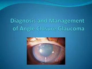
DEFINITION Angle-closure glaucoma is the glaucoma characterised by reduced aqueous outflow and elevated intraocular pressure due to blockade of trabecular meshwork by peripheral iris.
CLASSIFICATION OF ANGLE-CLOSURE GLAUCOMA Based On Pathogenic Mechanism A. WITH PUPILLARY BLOCK • Primary Angle-Closure Glaucoma • Acute • Sub acute • Chronic • SecondaryAngle-Closure Glaucoma • Swollen lens • Mobile lens syndrome • Miotic induced Continued:
B. WITHOUT PUPILLARY BLOCK • Primary Angle-Closure Glaucoma • Plateau iris configuration • Plateau iris syndrome • SecondaryAngle-Closure Glaucoma • Due to anterior pulling mechanism • Due to posterior pushing mechanism Continued:
Anterior Pulling mechanism • Neovascular Glaucoma • Iridocorneal endothelial syndrome • Posterior Polymorphous Dystrophy • Aniridia • Posterior pushing mechanism • Aqueous misdirection syndrome • Nanophthalmos • Cysts of iris and intraocular tumors • Intravitreal air injection • Suprachoroidal Hemorrhage • Scleral Buckling • Retrolental Fibroplasia
RISK FCTORS FOR DEVELOPMENT OF ANGLE-CLOSURE GLAUCOMA • AGE Common in old age i.e. 6th-7th decade of life. Reason: Continuous growth of lens Anterior displacement of lens Increased elasticity of iris Increased miosis • GENDER Females have three times higher incidence than males Reason: Females have shallower anterior chamber than males. Continued:
RACE Most common in South-East Asians, Chinese and Eskimos. Common in Caucasians. Least common in Blacks. • HEREDITY Most cases of primary angle-closure glaucoma are sporadic. No HLA association. Narrow angle characteristics are inherited under polygenic gene influence. • REFRACTIVE ERROR Common in hypermetropes; rare in myopes. Reason: Smaller eye and shallow anterior chamber in hypermetropes. Continued:
SEASON More common in winter months due to low illumination. • SYSTEMIC DISORDERS Inverse correlation between abnormal glucose tolerance and anterior chamber depth. • EMOTIONAL UPSET Due to excessive sympathetic activity. • DRUGS Sympathomimetics, anticholinergics and strong miotics.
PATHOGENESIS PRIMARY ANGLE-CLOSURE GLAUCOMA WITH PUPILLARY BLOCK Two factors are responsible: • Lens-iris apposition • Anatomic considerations
LENS-IRIS APPOSITION Contact of posterior iris surface with anterior lens surface Resistance to passage of aqueous from posterior to anterior chamber Relative pupillary block Greater pressure difference between posterior and anterior chambers Forward bowing of peripheral iris Blockade of trabecular meshwork Reduced aqueous outflow Rise in intraocular pressure Angle-Closure Glaucoma
ANATOMIC CONSIDERATIONS • Shallow anterior chamber (<2.5mm) • Decreased anterior chamber volume • Short axial length of globe • Small corneal diameter • Decreased corneal height • Increased posterior corneal curvature • Increased lens thickness • Anterior position of lens. • More anterior insertion of iris on ciliary body. • Increased curvature of anterior lens surface.
ROLE OF IRIS MUSCULATURE Forces Exerted By Iris Muscles: Parallel to the plane of iris Posteriorly SPHINCTER MUSCLE Posterior vector is: Minimum in miosis. Increases with dilatation. Maximum in mid-dilated(3-6mm)state. Crowding of angle by peripheral iris: Maximum in mid-dilated state. DILATOR MUSCLE Posterior vector of dilator muscle is more pronounced in a predisposed eye, i.e. an eye with shallow anterior chamber. During active dilation dilator muscle moves faster than the adjacent stroma, there by pulling the sphincter muscle closer to the lens and increasing the posterior vector of the latter.
SECONDARY ANGLE CLOSURE GLAUCOMA WITH PUPILLARY BLOCK Pupillary block occurs secondary to some pathological change in the eye. • SWOLLENLENS (PHACOMORPHIC GLAUCOMA) Swollen lensIris-lens apposition Pupillary block • MOBILE LENS SYNDROME (ECTOPIA LENTIS AND MICROSPHEROPHAKIA) Lens in anterior chamber Pupillary block • EXTREME MIOSIS (ANTICHOLINESTRASES) Pupillary constriction Lens-iris apposition Ciliary contraction Forward lens movement Pupillary Block
APHAKIA Adhesion of iris to anterior vitreous face Pupillary block • PSEUDOPHAKIA (ACIOL usually) Adhesion of iris to pseudophakos Pupillary block Secondary angle closure glaucoma with pupillary block due to dislocated PCIOL. Secondary angle closure glaucoma with pupillary block due to silicon oil.
PRIMARY ANGLE CLOSURE GLAUCOMA WITHOUT PUPILLARY BLOCK There is little or no pupillary block, still peripheral iris occludes the trabecular meshwork. PLATEAU IRIS CONFIGURATION Shaffer & Chandler Central anterior chamber depth : Normal Iris : Flat from pupillary margin to mid-periphery (plateau) Sharp turn posteriorly at mid-periphery and insertion at ciliary body creating a narrow angle recess. Glaucoma is cured by iridectomy. Associations : Anteriorly displaced ciliary body pressing on iris periphery. Ciliary body cysts.
PLATEAU IRIS SYNDROME Features are similar to Plateau Iris Configuration except that it is not cured by iridectomy.
SECONDARY ANGLE CLOSURE GLAUCOMA WITHOUT PUPILLARY BLOCK ANTERIOR PULLING MECHANISM • NEOVASCULAR GLAUCOMA Formation of ectropian uveae and latter peripheral anterior synechiae due to pull of the fibrovascular membrane over iris. • IRIDOCORNEAL ENDOTHELIAL SYNDROME Pull by the tonofilaments in the “epithelialised” endothelium and Descemet’s membrane of cornea over iris. • POSTERIOR POLYMORPHOUS DYSTROPHY Dysplastic corneal endothelium produces basement membrane like material which covers the angle.
POSTERIOR PUSHING MECHANISM • AQUEOUS MISDIRECTION SYNDROME Surgery/Insult to the eye Ciliary body swelling and forward rotation Contact with zonules/lens Aqueous secretion in vitreous pockets Anterior hyaloid and lens move forward Anterior chamber collapse. NANOPHTHALMOS Eye is normal in shape but smaller in size. Antero-posterior diameter < 20 mm. Corneal diameter < 11 mm Lens/Eye volume ratio :10-25% (Normal 3-4%) Angle closure is precipitated by choroidal effusion leading to forward rotation of ciliary body and loosening of zonules. • CENTRAL RETINAL VEIN OCCLUSION Decreased venous drainage of uveae Swelling and forward rotation of ciliary body Loosening of zonules Forward lens movement. Continued:
SUPRACHOROIDAL HEMORRHAGE • POSTERIOR SCLERITIS • SCLERAL BUCKLING • PANRETINAL PHOTOCOAGULATION • RETINOPATHY OF PREMATURITY.