Download
1 / 66
760 likes | 1.03k Views
Paediatrics teaching ppt. Immunodeficiency diseases. Xinhua Hospital Shanghai Institute for Pediatric Research Tong-Xin Chen. Development of Immune System. Development of IgG in Newborn Infant. Up to normal adults level From mother mainly

E N D
Paediatrics teaching ppt Immunodeficiency diseases Xinhua Hospital Shanghai Institute for Pediatric Research Tong-Xin Chen
Development of Immune System Development of IgG in Newborn Infant • Up to normal adults level • From mother mainly • Achieve to 60% of adults level when 1 year old,and 100% of adults level when 6 years old • IgG could be subdivided into IgG1、IgG2、IgG3 and IgG4 • Age dependent changes of serum IgG level synthesized by themselves:IgG1(5y);IgG3(10y);IgG2 and IgG4(14y)
Cord blood IgG level ≥ IgG from mother(>10% of IgG from mother ) • IgG from mother are catabolized gradually after born • IgG from mother disappeared completely when 6 months , serum IgG levels of 3~6 months infant are lowest ,especially IgG2 and IgG4
Development of IgM in Newborn Infant • IgM from mother can not pass placenta,serum IgM of fetuses synthesis when born <200-300mg/L • Normal neonatal IgM increase rapidly after born 4-7 days,is likely to be associated with the response of IgM to intestinal bacteria • If increasing,implicating neonates are stimulated by “nonself” antigen in uterus
Development of IgA in Newborn Infant • Can not pass placenta,serum IgA level achieve to 20% of adults level when 1 year old,and 100% of adults level when 12 years old • Cord blood IgA level ≤50mg/L,increasing of IgA implicates the possibility of infections in uterus • IgA is detectable in tears and saliva of 2-3 weeks neonate • The biological function of IgA is defend against some local mucous infections
Cellular Immunity of Newborn Infant • Number of T lymphocytes are usually normal • CD4+T cells are relatively higher,CD4/CD8 up to 3~4,consequently, are susceptible to infections • Function of Th2 cells are relatively stronger,are susceptible to allergic diseases • CD45RA+T cells are more,CD45RO+T cells are less • Deficiency of Cytokine :IFN-γ、IL-4,and so on
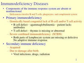
Immunodeficiency diseases,ID • A group of disorders characterized by an impaired ability to produce normal immune response. Most of these disorders are cased by mutations in genes involved in the development and function of immune organs, cells, and molecules. • Primary and acquired
Classification of Primary Immunodeficiency Diseases(7 main Categories) • Antibody or B cells deficiency(50%) • Combined immunodeficiency(20%) • Phagocytic dysfunction(18%) • Cellular or T cell deficiency (10%) • Complement deficiency(2%) • Immunodeficiency with other important characteristics • Immunodeficiency with or acquired other congenital or hereditary diseases
The incidence of PID • Calculated by alive infants:1/10000 (Japan 1981 and Australia 1983) • Hongkong report:1/8000 • There is no statistics report in mainland so far • According to the incidence of 1/10000,2500/25000000 nerborn infants every year in our country,cases add up to 3~8 ten thousands at least • More than 100 cases in our hospital since 1970
Clinical features • Recurrent infection • High risk of autoimmune diseases • High risk of malignancy
Infection • Severe infection、 • Refractory infection、 • Recurrent infection、 • Opportunistic pathogens infection、 • Recurrent diarrhea、
Children with immunodeficiency have higher risk of autoimmune than normal(0.01%~14%) Autoimmunity diseases Autoimmune disease suspicious Arthritis SLE,JRA Thrombocytopenia ITP Neutropenia Crohn’s desease SLE Hemolytic anemia Immunodeficiency associated with autoimmune X-Linked Agammaglobulinemia Selective IgA Deficiency CVID Thymic hypoplasia Immunodeficiency with hyperIgM Chronic granulomatosis Complement deficiency Wiskott-Aldrich Syndrome
Children with immunodeficiency have higher risk of cancer than normal(100~300 fold) Tumor
Primary immunodeficiency suspicious History • Tympanitis more than 8 times per year • Severe nose sinusitis more than 2 times per year • Pneumonia more than 2 times per year • Deep infection in abnormal position more than 2 times • Recurrent infection in deep skin or viscera • Infection eliminated with antibiotics by intravenous injection • Rare or opportunistic pathogens infection • Family PID history
Clinical features • Growth development deficiency • Lymph nodes or tonsil deficiency • Skin changes:capillary vessel expand, petechia • Skin mildew, lupus erythematosus-like tetter • Ataxia(A-T) • Thrush after 1 year old • Oral ulear
Laboratory analysis: • Serum IgG,IgM,IgA(B cell function) • CD3,CD4,CD8 (T cell subsets) • CD19( number of B cell ) • CD56/16( NK cell) • White blood cell count or nitroblue tetrazolium( NBT )test • Complement
IgG subclasses(1~4) • Thymus:X -ray • Cytokine: IL-2,IL2R,IFN,IFNR • Cell surface molecular:CD18 • Gene analysis:BTK ,CD40L,WASP
Common primary immunodeficiency diseases • Combined immunodeficiency(14) • Antibody or B cells deficiency(10) • Cellular or T cell deficiency(9) • Immunodeficiency with other important characteristics(3) • Phagocytic dysfunction(12) • Complement deficiency(16) • Immunodeficiency with or acquired other congenital or hereditary diseases(41)
X-linked agammaglobulinaemia • Selective IgA deficiency • Thymic hypoplasia • Combined immunodeficiency
X-linked agammaglobulinaemia ( XLA ) • Also named as Bruton disease(described in 1952) • Discovered that the disease was associated with mutation of the gene coding pre B-cell cytoplasmic tyrosine kinase( btk)in1993 • Mutation lead to block in signal transduction of B cell development,block in maturation after the pre-B cell stage ,lead to decreaseing of mature B cell • The patterns of mutations are diverse,more than 118cases are reported so far
Clinical features • Male • Onset during 4~6 months of age • Recurrent Pyogenic bacterial infection • Respiratorty tract infections are typical,as well as systemic infections
Immunological characteristics: • Can hardly produce antibody,all kinds of Ig are markedly reduced • IgG < 2g/L ( <200mg/dl ) • IgA <0.02g/L ( <2mg/dl ) • IgM <0.1g/L ( <10mg/dl ) • Circulating B cells are markedly decreased,usually less than 0.5% of total lymphocytes • Numbers and function of T lymphocytes are normal • Btk gene located on Xq21.3-22 is deficiency
Mutation of Btk gene 62145-62155 cDNA mutation: 989_999delTGACTCGGAGTinsGGTGGTATTCCAAA Codon change: MTRS286_289RWYSK Mother status: carrier Exon10F 62155-62145 Exon10R
Differential diagnosis : infantile transient hypogammaglobulinemia 1~2y normal reduced normal unclear presence no Characteristics Age IgM IgG IgA Molecular deficiency B cell IgGreplacement? XLA congenital(>6m) reduced absent/ reduced absent/ reduced BTK absent/ reduced yes
Common Variable Immunodeficiency ( CVID) • A heterogeneous group of diseases characterized by antibody defects • Late-appearing immunodeficiency • Immunological characteristics of CVID • Antibody deficiency IgG <2.5g/L ( <250mg/dl ) • IgA usually is reduced • IgM usually is reduced • Circulating B cells usually are normal or decreased • Cellular immunity:normal or help function deficiency
Clinical manifestations: • Recurrent infection ( bacterial infection)with onset at any age, affects both males and females • High risk of gastrointestinal infections,usually chronic giardiasis • Lymphoma and gastric carcinoma occur with increased frequency • Increased incidence of autoimmune disease(hemolytic anemia 、pernicious anemia、 thrombocytopenia,et al) • lymphoproliferation,splenomegalia,lymphoid hyperplasia
X-linked agammaglobulinaemia • Selective IgA deficiency • Thymic hypoplasia • Combined immunodeficiency
Incidence:Caucasian1/500~1500,Japanese1/18500,Chinese1/5000~10000Incidence:Caucasian1/500~1500,Japanese1/18500,Chinese1/5000~10000 • Associated with maladjustment of Th2 cell to B cell produce IgA • Both males and females, often coincide in same family • Mild form is asymptomatic • Recurrent infections in infancy(respiratory 、intestinal and urinary infections ) • Be associated with autoimmune diseases、asthma and intestinal malabsorption
Serum IgA less than 0.05g/L,IgM、IgG normal or increased • sIgA markedly reduced • Serum IgA could increase to normal level in some cases • Should not be treated with IVIG,since capable of forming anti-IgA antibodys subsequent allergy
X-linked agammaglobulinaemia • Selective IgA deficiency • Thymic hypoplasia • Combined immunodeficiency
Thymic hypoplasia also is called DiGeorge syndrome(1964年) • It is known now that 80%~90% Digeorge syndrome have minor deletion of gene located at 22q11 • Minor deletion of gene located at 22q11 included a group of disease,now called CATCH22 syndrome
CATCH 22 Cardiac defects Abnormal facies Thymus hypoplasia Cleft palate Hypocalcemia
Clinical features: Thymus hypoplasia Ⅲ-Ⅳpharyngeal archhypoplasia Ⅰ-Ⅱpharyngeal archhypoplasia Parathyroid hypoplasia Abnormal facies Cardiac defects T cell reduced Hypocalcemia Recurrent infections(virus infections ) Cleft palate、short philtrum and low-set ears Tetralogy of Fallot and aorta abnormal(eg.arcus aortae break off) Tetany
Laboratory analysis: • Number of peripheral blood lymphocytes reduced(<1000个/mm2) • CD3+T cell markedly reduced • Serum Ig normal or reduced,whereas IgE increased • Serum calcium reduced, serum phosphorus increased, parathyroidhormone reduced • Chest radiograph: thymus absence
DiGeorge syndrome: Boy 14months Bronchopneumonia Congenital heart disease Immunodeficiency Hypocalcemia Nearside facial paralysis
Normal Thymus Thymus shadow of infant(<6m) is visible ,usually>10g If invisible(< 4g ),implicated thymushypoplasia
DiGeorge syndrome Thymus absence
X-linked agammaglobulinaemia • Selective IgA deficiency • Thymic hypoplasia • Combined immunodeficiency
A group of diseases,occurs both males and females in autosomal recessive SCID,only males in X-linked recessive SCID • Recurrent infections with fungi, bacteria, virus, mycobacterium, protozoa • Typical features: chronic diarrhea、pneumonia and persistent fungal infections(especially thrush) • Sometimes morbilliform rash is the only symptom of SCID in neonatal period ,may caused by GVHR • Usually succumb to overwhelming infection whithin the first year of life
Severe combined immunodeficiency (SCID) T- B+NK-Ig- • Approximately 50%~60% SCID are X-linked forms ,the most common genetics alteration is mutation of receptor c of IL-2、IL-4、IL-7、IL-9 and IL-15 • Autosomal recessive SCID usually have JAK3 gene deficiency,JAK3 coded a tyrosine protein kinase which is associated with signal transduction initiated by c
T- B-NK+Ig- • Autosomal recessive SCID may have mutations of RAG-1 and RAG-2,affects antigen receptor on T、B cells surface • In addition, approximately 50% autosomal recessive SCID have adenosine deaminase (ADA)deficiency
Boy ,8months Recurrent pneumonia、thrush One of his uncle died at 6months unknown cause T-B+NK-Ig ↓ Figure 8 photo of a patient with SCID : candida albicans in the mouth
Boy ,4.5months Fever ,pneumonia,hepatosplenomegaly ,Have abscess after inoculating BCG vaccine 3 months ,rash and diarrhea after transfusion Figure 8-2 photo of a patient with SCID : GVHD and BCG vaccination
Molecular Diagnosis of X-SCID in Patient 1 IL2RG gene PCR direct sequencing 487bp deletion • Deletion mutation from intron 6 to 7 including exon 7 and 2 primer site (IVS6-71 to IVS7-11del487) • Predicted frameshift start at arginine 285 with stop codon (TAA)created at position 342, predicted premature termination (R285fsX342) Patient 1: deletion between Intron 6 and intron 7 Deletion in patient Deletion in patient IVS6-17 IVS7-11 Normal control: Intron 6 Normal control: Intron 7
-ve control -ve control Patient Patient Mother Mother Normal Normal Primer Pair Exon 6F/8R Primer Pair Exon 5F/8R Carrier diagnosis in IL2RG deletion (XSCID) – Patient 1PCR-agarose gel electrophoresis Causative gene: IL2RG in X-chromosome • PCR amplified for each exon for sequencing • No PCR product for amplification of exon 6, 7 and 8 • Suspected large deletion, try other primer pairs combination • Deletion mutation including exon 7 and 2 primer site found (IVS6-71 to IVS7-11del487) • Mother diagnosed as heterozygous carrier by PCR directly
Hyper IgM syndrome (HIGM) T+CD40L-B+IgM↑IgG↓ • Four types,most common type is X-linked form (Hyper IgM syndrome typeⅠ) • Approximately 70%,caused by mutations of the CD40L gene • Diagnosis: CD40L expression on T cell reduced in vitro lymphocyte cultivation