Download
1 / 12
150 likes | 359 Views
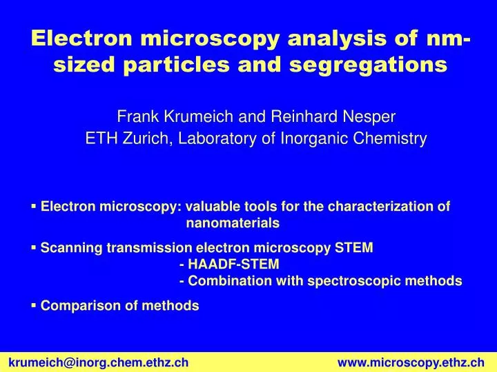
Electron microscopy analysis of nm-sized particles and segregations. Electron microscopy: valuable tools for the characterization of nanomaterials

E N D
Electron microscopy analysis of nm-sized particles and segregations • Electron microscopy: valuable tools for the characterization of nanomaterials • Scanning transmission electron microscopy STEM - HAADF-STEM - Combination with spectroscopic methods • Comparison of methods Frank Krumeich and Reinhard Nesper ETH Zurich, Laboratory of Inorganic Chemistry krumeich@inorg.chem.ethz.chwww.microscopy.ethz.ch
Electron Microscopy Methods for the Characterization of Nanomaterials (Example: Vanadium Oxide Nanotubes) SEM: characterization of tubularmorphology EELS: composition V map C map Cross-sections of VOx nanotubes: TEM and elemental maps obtained by electron spectroscopic imaging TEM: characterization of the wall structure
Scanning Transmission Electron Microscopy (STEM) STEM detectors HAADFHigh Angle Annular Dark Field detector (Θ > 3°) BFBright Field detector ADFAnnular Dark Field detector (Θ = 0.5 - 3°)
Scattering of Electrons at an Atom Strong Coulomb interaction of an electron with the nucleus scattering into high angles or even backwards High angle annular dark field detector (HAADF-STEM) atomic-number (Z) contrast:
10 nm HAADF-STEM of Small Metal Particles 50 nm Au particles (bright contrast) on titania (Z contrast)
Al C O Pt Pd Cu Pt Pt Al C O Cu Pt Pd Pt Pt HAADF-STEM and EDXS: Point Analyses Pd/Pt particles on alumina
matrix segregation HAADF-STEM and EDXS WO3 segregations in the oxidation product of Nb4W13O47 (Tox=1000°C) Krumeich, Nesper, J. Solid State Chem.179 (2006) 1658
100% W ca. 80% Nb Single-crystal X-ray structure of Nb7W10O47 P21212 a=12.26, b=36.63, c=3.95 Å (Krumeich, Wörle, Hussain, J. Solid State Chem.149 (2000) 428) HAADF-STEM: Elemental Distribution HAADF-STEM of Nb4W13O49
High-Resolution Electron Microscopy HAADF-STEM HRTEM WO3 segregations in a bronze-type Nb-W oxide 2 nm
Analytical Electron Microscopy Benefits • Qualitative and quantitative information about the composition: EDXS, EELS • Bonding, coordination, interatomic distances: Fine structure in EELS (ELNES, EXEFS) • Spatially resolved information about composition:1. STEM + EDXS and/or EELS2. ESI • Electron-matter interactions are mostly elastic high electron doses necessary • Long measuring times high sample stability and absence of drift • Ionization edges occur at different energies and are of different shape not all methods are equally suitable for all elements Limitations
Post-column filter Transmission Electron Microscope Tecnai F30 Uacc= 300kV, field emission cathode (FEG) SuperTwin lens: Cs = 1.15 mm, point resolution d < 0.2 nm Equipment: post-column imaging filter, STEM, energy-dispersive X-ray spectrometer Methods: TEM, HRTEM, STEM, ED, EDXS, EELS, ESI, EFTEM Acknowledgements EMEZ: Electron Microscopy Center, ETH Hönggerberg www.emez.ethz.ch krumeich@inorg.chem.ethz.chwww.microscopy.ethz.ch