Download
1 / 28
680 likes | 3.42k Views
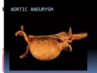
Sinus Valsalva Aneurysm. Seoul National University Hospital Department of Thoracic & Cardiovascular Surgery. Sinus Valsalva Aneurysm. Definition Thin walled, saccular or tubular outpouchings, usually always in the right sinus or adjacent half of the noncoronary sinus.

E N D
Sinus Valsalva Aneurysm Seoul National University Hospital Department of Thoracic & Cardiovascular Surgery
Sinus Valsalva Aneurysm • Definition • Thin walled, saccular or tubular outpouchings, usually always in the right sinus or adjacent half of the noncoronary sinus. • They generally have an intracardiac course, but may protrude into the pericardial space and they may rupture into the right (or rarely left) heart chambers to form an aorta-cardiac fistula. • This defect may result from absence of normal elastic tissue and media in this region. • Congenitally weak area gradually enlarges under aortic pressure to form an aneurysm, although the age at which this occurs is uncertain
Sinus Valsalva Aneurysm • History • 1st description by Hope in 1839 • 1st important paper published by Thurman in 1840 • Suggested ruptured as congenital by Abbott in 1919 • Reviewed the subject of congenital and acquired lesion by Jones and Langley in 1949 • 1st diagnosis of rupture during life by Venning in 1951 • 1st. successful repair with CPB in 1956 at Mayo Clinic & University of Minnesota using CPB
Aneurysm of Sinus Valsalva • Clinical features • Etiology ; congenital but other possibly acquired • * Endocarditis, syphilis, Behcet’s disease, atherosclerosis, • Cystic medial necrosis, penetrating injury • * Incomplete fusion of proximal & distal bulbous chordous • * Anatomic defect in the elastic tissue • * Deficiency of the conal septum • Rupture or fistula • 1) Incidence : rare ( 0.2 ~ 0.5% of open heart surgery), • 75% ~ 80% are male • 2) Site • * Right coronary sinus to right ventricle : 65% • * Noncoronary sinus to right atrium : 25% • * Left coronary sinus to left atrium : rarely • 3) Aortico-left ventricle tunnel • : exceedingly rare form
Sinus Valsalva Aneurysm • Etiology • Separation of the aortic media of the sinus from the media adjacent • to the hinge line of the AV valve cusp resulted from the absence • of normal aortic elastic tissue and media in two region. • Congenitally weak area gradually gives way under aortic pressure to • form an aneurysm. • The aneurysm appears an excavation of the sinus which protrudes • into the underlying cardiac chamber. • In Asians, the basic abnormality is sited leftward and toward the • commissural area between Rt. and Lt. cusp. • Acquired lesions caused by medionecrosis, syphilis, atherosclerosis, • endocarditis, or penetrating injury are more diffuse, involving more • of sinus or multiple and often ascending aorta, and projecting outside • the heart.
Sinus Valsalva Aneurysm • Pathophysiology • Thinning of the aorta medial layer in the wall of a sinus of Valsalva results in an aneurysmal dilation, which may extend and rupture into a corresponding cardiac chamber, forming an aortocardiac fistula. • Aneurysms usually arise from the right coronary sinus and extend into the right ventricle or right atrium. • Aneurysmal rupture into the right heart results in a large left-to-right shunt , which, in turn, can lead to congestive heart failure. • Unruptured aneurysms extending into the right heart may cause tricuspid valve stenosis/incompetence, right ventricular outflow tract obstruction, or complete heart block.
Sinus Valsalva Aneurysm • Histologic view Unruptured aneurysm of right sinus Valsalva Aneurysm is walled by atrophic muscular tissue of RVOT
Sinus Valsalva Aneurysm • Associated cardiac anomalies • VSD occurs in 30 to 50%, but may be a little higher in surgical patients. • Aortic valve abnormalities & incompetence are common, and when VSD is present, AR usually results from a prolapsed cusp , and when VSD is not present , AR usually arises from other valve abnormalities • Pulmonary stenosis is uncommon, but small gradients are common. • Others are uncommonly, but any defects including COA, PDA, ASD, subaortic stenosis & TOF are present.
Sinus Valsalva Aneurysm • Natural History • Unruptured aneurysms uncommonly cause symptoms, by • protrusion into RA and RV, heart block as well as ventricular • tachycardia may result. • Rupture of aneurysm tends to take place in the 3rd or 4th • decade of life. • Once symptoms develop, the heart failure worsens and, without • surgical treatment, most patient die within one year. • Clinical presentation is usually within the 3rd decade of life • When a VSD coexists, AV is usually at least mildly incompetent, • by the time 15 to 20 years, a fixed fibrous deformity of the • prolapsed leaflet occurs.
Sinus Valsalva Aneurysm • Clinical features • The SVA produce TV dysfunction or RVOT obstruction. • 80% of the persons with sinus Valsalva aneurysm are male. • Rupture produces acute symptoms in about 35% and gradual onset of effort dyspnea in 45% and no symptoms in 20%. • In a few patients, death occurs within days, but in most there is improvement, followed by recurrent symptoms. • The frequency of symptoms may be related to the size of the Fistula. • Rupture is heralded not only by pain & dyspnea but also by appearance of murmur, widened pulse pressure.
Sinus Valsalva Aneurysm • Daigram of aneurysm • Unruptured aneurysm of right sinus Valsalva with VSD
Sinus Valsalva Aneurysm • Daigram of aneurysm • Ruptured right Sinus Valsalva Aneurysm with VSD
Sinus Valsalva Aneurysm • Rupture site Arrows indicate common sites of rupture of sinus of Valsalva aneurysm M ; membraneous septum NC ; noncoronary sinus V ; atrioventricular septum C ; conal septum
Sinus Valsalva Aneurysm • RVOT obstruction
Sinus Valsalva Aneurysm • Rupture • The sinus of origin is the main determinant of the direction of projection • and rupture. • Gradually develops a more localized windsock, in an unknown • percent of cases ultimately rupture into an adjacent low pressure • chamber and rarely outside chamber. • When the aneurysm coexists with a VSD(30-50%), the windsock usually projects into the RV. • In about one fourth, there is no windsock or any suggestion of • aneurysm formation, but rather, a direct fistulous communication. • Typical windsock deformity may be more common from right sinus • lesion, and a direct fistula in noncoronary sinus to RA lesion. • .
Sinus Valsalva Aneurysm • Sites of rupture or fistula • Aneurysm of the right sinus may originate more centrally and project into the outlet of RV, but leftward portion into region of • membranous septum. • Aneurysms from the noncoronary sinus usually originate from its anterior portion and rupture into the RA, but in rare cases • into RV, posterior portion may rupture into the pericardium. • Rarely, right or noncoronary sinus aneurysm rupture into LV. • Aneurysms from left coronary sinus rupture into the LA, LV, but rarely into LV due to thick wall and high pressure. • Aneurysms rupturing into areas adjacent to TV may be a cause of heart block or RBBB.
Sinus Valsalva Aneurysm • Indications for operation • When ruptured or is associated with VSD or with a VSD and AR, prompt operation is advisable. • Unruptured aneurysm that are producing • hemodynamic derangements should be repaired. • Small or moderate-sized unruptured aneurysm • probably should not be repaired surgically.
Sinus Valsalva Aneurysm • Techniques of operation • Ruptured aneurysm of right sinus Valsalva without • VSD • Ruptured aneurysm of the sinus of Valsalva into the • RA without VSD • Ruptured aneurysm of the right sinus of Valsalva • associated with VSD • * Repair by excision of aneurysm and reconstruction • * Repair by closing the origin of aneurysm • * Repair the associated VSD and valve
Sinus Valsalva Aneurysm Techniques of operation • Repair of ruptured aneurysm of right sinus Valsalva with VSD
Sinus Valsalva Aneurysm Techniques of operation • Repair of unruptured aneurysm of right sinus Valsalva
Sinus Valsalva Aneurysm Techniques of operation • Noncoronary sinus of Valsalva aneurysm extending into the right atrium
Sinus Valsalva Aneurysm Techniques of operation • Noncoronary sinus of Valsalva aneurysm extending into the right atrium • VSD patch closure in case of VSD
Sinus Valsalva Aneurysm • Techniques of operation • David-V valve-sparing root replacement using a De Paulis • Gelweave Valsalva graft
Sinus Valsalva Aneurysm • Results of operation • Survival • Risk factors for premature late death • 1) severe aortic incompetence • 2) left ventricular enlargement • 3) aortic valve replacement • Functional status • Persistent or worsening aortic valve incompetence • accounts for most of functional disability • Complications • 1) Reoperation • 2) Heart block
Aorta–right Atrial Tunnel • Clinical features • Aorta–right atrial tunnel (ARAT) is a very rare abnormal tubularextracardiac communication between the ascending aorta and theright atrium. • The first case wasdescribed in 1980 by Otero Coto and colleagues • Embryologic background and cause for this anomaly are not clear. • Probable cause seems to be a congenital deficiency of the elastic lamina inthe aortic media • The tunnel-like vascular extracardiac communication betweenthe aortic root and the right atrium arose from any of the 3sinuses of Valsalva.
Aorta–right Atrial Tunnel • Clinical features • This aorto–right atrial communication behaves like a left-to-rightshunt at the atrial level. • The most common symptoms were shortness of breath, palpitation,and recurrent respiratory tract infections. • On physical examination,all patients had a continuous murmur at the right parasternalborder. • The single diagnostic feature is demonstration of this distincttunnel arising from one of the aortic sinuses of Valsalva andhaving an extracardiac course and entering into the right atrium • Treatment options are simple ligation or ligation with implantationof coronary ostium or coil embolization.