Download
1 / 20
210 likes | 428 Views
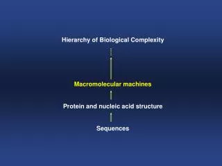
Macromolecular Machines - Introduction. Biochemistry 4000 Dr. Ute Kothe. Macromolecular Machines. DNA Polymerase – DNA replication, Voet chapter 30 error rate: 10 -7 2. RNA-Polymerase – Transcription, Voet chapter 31 error rate: 10 -3 3. Ribosome – Translation, Voet chapter 32

E N D
Macromolecular Machines- Introduction Biochemistry 4000 Dr. Ute Kothe
Macromolecular Machines • DNA Polymerase – DNA replication, Voet chapter 30 • error rate: 10-7 • 2. RNA-Polymerase – Transcription, Voet chapter 31 • error rate: 10-3 • 3. Ribosome – Translation, Voet chapter 32 • error rate: 10-3 - 10-4 Unifying question: How do all these machines achieve high accuracy in duplication and expression of genetic information? In each case, Watson-Crick base-pairs have to be selected with high accuracy against non-Watson-Crick base pairs.
The Ribosome Large Subunit (50s) Small Subunit (30s) Decoding center E-site P-site A-site tRNAs Voet, Fig. 32-36
E. coli Ribosome composition Voet, Table 32-7
Translation Elongation Cycle 1. Decoding / A-site binding 3. Translocation 2. Peptide bond formation Voet, Fig. 32-48
Ternary complex EF-Tu-GTP-aa-tRNA EF-Tu as aG protein switch EF-Tu-GTP • EF-Tu-GTP: • on/active • can bind aa-tRNA • EF-Tu-GDP: • off/inactive • can not bind aa-tRNA due to large scale domain rearrangements • EF-Ts: • Guanine nucleotide exchange factor EF-Tu-GDP EF-Tu-EF-Ts
Cryo-EM: Ribosome + EF-Tu-GTP-aa-tRNA Fitting of the crystal structures of ribosome and EF-Tu-GTP-aa-tRNA into the electron density 13 Å resolution Stark et al., NSB 2002
Ribosomal Decoding site Upon binding of the correct tRNA, A1492 and A1493 flip out and interact with the codon-anticodon duplex. Voet, Fig. 32-64
1st position: A1493 anticodon codon The decoding site: shape recognition 3rd position: G530 2nd position: A1492 anticodon codon anticodon codon 1st and 2nd position monitor geometry of Watson-Crick basepair by measuring Distances between riboses! No specific interaction with bases! Relaxed monitoring of 3rd codon position Decoding Problem: difference in binding energies of cognate versus near-cognate (one mismatch) tRNAs not sufficient for efficient discrimination Voet, Fig. 32-63
DNA Polymerase I • bacterial DNA-Polymerase, single polypeptide, highly processive • Proofreading ability: 3’-5’ exonuclease, 5’-3’ exonuclease • Klenow fragment: C-terminal fragment with polymerase & 3’-5’exonuclease activity Voet, Fig. 30-8
Taq Polymerase +/- substrate NTP Voet, Fig. 30-9 Closed conformation Incoming nucleotide bound O helix (orange) closes over active site Open conformation No incoming nucleotide O helix away from active site
Recognition of incoming dNTP • Recognition of shape of base pair independent of hydrogen bonding properties • Conserved Tyr stacks on template base • Last 3 nucleotides in A-DNA conformation with wider minor groove which is monitored by amino acids for N3 of purines and O2 of pyrimidines • 2,4-Difluorotoluene (F) can be inserted instead of thymine (T) by DNA-Pol I (isosteric, but can not accept hydrogen bonds)
Catalytic Mechanism of DNA-Pol. • Most likely common catalytic mechanism for all DNA-Pol.: • metal ion A (Mg2+) acitvates 3’OH of primer for nucleophilic attack on a-phosphate • metal ion B (Mg2+) orients triposphate group for in-line attack and shields negative charges as well as additional charges in transition state Voet, Fig. 30-10
Animal DNA Polymerases Voet, Table 30-5
RNA-Polymerase Bacteria: a2bb’ws Voet, Table 31-2
Taq RNA-Polymerase Holoenzyme with s subunit a2bb’w – core enzyme a – yellow & green b – cyan b’- pink w - gray Voet, Fig. 31-11 & 12
Yeast RNA-Polymerase II Note similarity to bacterial RNA-Polymerase! View from the right in left part Voet, Fig 31-20
RNA-Pol II Elongation complex • “clamp” swings over DNA to trap it, ensures high processivity • unwound template strand make 90° turn after active site due to “wall” • active site accessible through funnel for new NTPs • sequence-independent contacts of enzyme with sugar-phosphate backbone • DNA-RNA hybrid helix is disrupted by “rudder” Voet, Fig. 31-21
Transcription cycle bent • Highly conserved “bridge helix” contects two pincers forming the enzyme’s cleft • Bridge helix nonspecifically contacts template DNA at +1 position • Straight in RNA-Pol II, bent in Taq RNA-Pol. • Might alternate between straight and bent conformation moving by 3- 4 A • Might push paired nucleotide at position +1 to position -1 during translocation Voet, Fig. 31-22
NucleotideAdditionCycle T7 RNA Polymerase Steitz, EMBO 2006