Download
1 / 30
320 likes | 539 Views
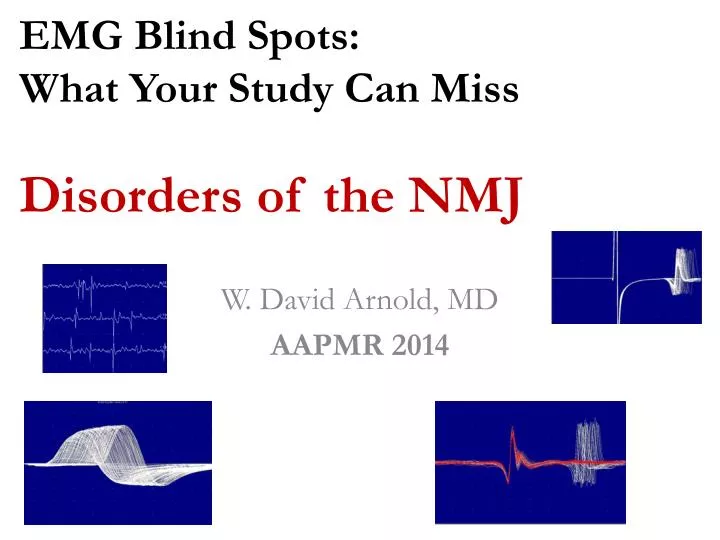
EMG Blind Spots: What Your Study Can Miss Disorders of the NMJ. W. David Arnold, MD AAPMR 2014. Outline. Review of Prototypical NMJ Disorder, Myasthenia Gravis, and the General Approach to NMJ Disorders

E N D
EMG Blind Spots: What Your Study Can Miss Disorders of the NMJ W. David Arnold, MD AAPMR 2014
Outline • Review of Prototypical NMJ Disorder, Myasthenia Gravis, and the General Approach to NMJ Disorders • Review a series of 4 Cases to Highlight Potential Pitfalls and Key Concepts in NMJ Edx studies
Case 1: “Proximal Limb Weakness” • 18 year old woman presents with a progressive proximal limb weakness is sent to the EMG Lab Limb Girdle Muscular Dystrophy? • Examination: • 4 grade proximal limb weakness • Normal bulbar strength, sensation, and reflexes
Case 1: NCS and EMG • Normal median sensory and motor response • EMG: short duration, small amplitude motor unit action potentials with early recruitment without fibrillations
Case 1: Proximal Limb Weakness (Cont.) • EMG and NCS: Non-irritable myopathy • Diagnostic consideration was given to a possible limb girdle muscular dystrophy • Muscle Biopsy: normal • Thoughts?
Case 1: Additional Studies • Repetitive ulnar nerve stimulation at 3 Hz ~50% decrement • SFEMG Increased jitter and blocking
Follow up Lab testing: • Elevated acetylcholine receptor antibodies Diagnosis: acetylocholine receptor antibody positive myasthenia gravis (MG) Treatment: Excellent response to pyridostigmine and immunomodulatory treatments
Clinical Pearl of Case 1: Ask the Correct Question 338 charts of patients with NMJ disorders 6 patients underwent muscle biopsy for possible myopathy 2 with coexistent inflammatory muscle dz 4 with NMJ disorders mimicking myopathy (based on normal muscle bx) Consider RNS in patients with weakness
MG • MG is the most common NMJ disorder • Prevalence of 20 in 100,000 and incidence of 2 per 100,000. (Phillips Ann N Y Acad Sci. 2003) • Antibodies • Ach receptor antibodies (80%) • Muscle specific tyrosine kinase (MuSK) (10%) • Antibody negative (5%-10%) • Treated with cholinesterase inhibitors and immunomodulatory treatments
NMJ Disorders: History • Weakness: proximal muscles with fatigability • Severity of involvement: Ocular>bulbar>limb>respiratory • Sensory: No sensory loss, paresthesia, or neuropathic pain • Autonomic: dry mouth, blurry vision, impotence, constipation, and difficulty with urination • Some disorders such as Lambert Eaton myasthenic syndrome (LEMS) and botulism Generalized fatigue ≠ NMJ disorder
NMJ Disorders: Examination • Weakness in proximal limb, ocular, and bulbar muscles • Distal or focal weakness may occur in 12% of MG (Wirtz et al 2002) • Extraocular and bulbar weakness are early and prominent in MG and botulism • Less prominent extraocular weakness in LEMS • Reflexes are normal in postsynaptic disorders such as myasthenia gravis but characteristically absent or reduced in presynaptic NMJ d/o • Sensory function is normal • Pupillary responses are usually reduced in botulism
Post-synaptic NMJ disorders • Sensory NCS -normal • Motor NCS -usually normal CMAP amplitude • EMG -Possibly short duration, low amplitude, unstable MUAP’s if severe (Jiggle) • Slow RNS (3 Hz) -decrement (60-80% of generalized MG) • CMAP with 10 sec exercise (or Rapid RNS, 20-50 Hz) -no increment • CMAP with Exercise -no increment • SFEMG -Jitter and blocking (95-98% MG) (Nl jitter in weak muscle ≠ MG)
Pre-synaptic NMJ disorder Sensory NCS -normal Motor NCS -usually low CMAP amplitude EMG -Possibly short duration, low amplitude, unstable MUAP’s if severe -some disorders with fibrillations (botulism) Slow RNS (3 Hz) -decrement CMAP with 10 sec exercise (or Rapid RNS, 20-50 Hz) -increment 300% in one muscle or 100% in three (diagnostic for Lambert Eaton MS) SFEMG -Jitter and blocking (jitter and blocking decrease with increasing firing rate)
Presynaptic NMJ Pre-exercise Post-exercise 350% increment following 10 seconds exercise
Case 2: Dysarthria • 64 year old woman with symptoms of progressive dysarthria and facial weakness is sent to the EMG Lab Possible myasthenia gravis? • Examination • Mild facial and moderate tongue weakness • Tongue fasciculations • Normal limb strength • Generalized Brisk reflexes and jaw jerk
Case 2: Nerve Conduction Studies Median and ulnar sensory responses are normal Median and ulnar motor responses are normal Repetitive nerve stimulation at 3 Hz -Orbicularis Oris with >20% decrement • Myasthenia Gravis?
Case 2: Electromyography Single Fiber EMG -Increased jitter and blocking in frontalis and extensor digitorum EMG Fibrillations/fasciculations/dec recruitment/enlarged motor unit action potentials (mentalis and tongue) Limb muscles normal Jitter and Blocking
Clinical Pearl of Case 2: Not all that Jitters is Myastheniathe “False Positive” test Amyotrophic Lateral Sclerosis & NMJ transmission defect • Severe decrement frequently seen on RNS (reported up to 35% of CMAP amplitude) (I have seen up to 50%) • Increased decrement correlates with faster progression and worse prognosis Daube 2004; Wang 2001 • SFEMG: increased jitter and block may be early finding, prior to denervation (also seen in early reinnervation) • Axonal Stimulation SFEMG: lack of facilitation with increasing frequency of stimulation Arimura 1995
Case 3: Seronegative MG A 58 year old woman with a 30 year history of seronegative myasthenia gravis PMH: poliomyelitis at age 6 requiring ventilation support in an iron lung for a month PE: normal CN exam, severe proximal limb weakness with fatigability, normal sensation and reflexes
Case 3: Seronegative MG • 3Hz RNS with up to 70% decrement in proximal muscles • She describes dramatic improvement with pyridostigmine • Worsening with prednisone • No improvement with IVIG or PLEX • Ideas?
Clinical Pearl of Case 3:Hoof beats (aka decrement)? Don’t assume it is a horse (MG) • Multiple NMJ disorders may fall within this clinical presentation • Seronegative (autoimmune) MG • Congenital Myasthenic syndromes • Other neuromuscular disorders with secondary NMJ effects or clinical patterns similar to NMJ disorders
Congenital Myasthenic Syndrome • Historical points in case 3 suggesting this possibility: • No clear response to immunomodulation • Atypical phenotype (Severe disease with minimal to no bulbar involvement) • PMH of “polio” after which she states she was “normal” in every way until developing weakness in her 20’s • On more direct questions she described being a “blue baby” at birth • Electrodiagnosis of CMS
Case 4: MG second opinion 45 year old man with progressive ptosis, dysphagia FH: mother with “seronegative myasthenia gravis” Examination: bilateral ptosis and extraocular muscle weakness without clear fatigability; mild proximal limb weakness Antibody testing: AChR and MuSK Antibodies (-) EMG and NCS: decrement on repetitive nerve stimulation studies Diagnosed with seronegative myasthenia gravis but then had minimal response to immune therapies and pyridostigmine? Ideas?
Case 4: Repeat EMG and NCS • NCS: multiple mononeuropathies c/w the patient’s known HNPP • RNS: normal • SFEMG: mildly increased jitter in frontalis with no blocking • EMG: Short duration small amplitude motor unit action potentials with sparse fibrillations in proximal limb and facial muscles
Clinical Pearl of Case 4: When in doubt, trust your exam… double check technique Movement artifact is significant challenge in repetitive nerve stimulation and can lead to false positive tests Our patient: • Lacked clinical features of fatigability or response to treatment • Diagnosed with oculopharyngeal muscular dystrophy (OPMD) • Autosomal dominant slowly progressive hereditary muscle disorder • GCG repeat expansion (8-13) • Often mistaken for seronegative MG • Typically later/less involvement of extraocular muscles
Thanks! Questions? William.arnold@osumc.edu
References • Phillips LH, 2nd. The epidemiology of myasthenia gravis. Annals of the New York Academy of Sciences. Sep 2003;998:407-412. • WirtzPW, van Dijk JG, van Doorn PA, et al. The epidemiology of the Lambert-Eaton myasthenic syndrome in the Netherlands. Neurology. Jul 27 2004;63(2):397-398. • WirtzPW, Sotodeh M, Nijnuis M, et al. Difference in distribution of muscle weakness between myasthenia gravis and the Lambert-Eaton myasthenic syndrome. Journal of neurology, neurosurgery, and psychiatry. Dec 2002;73(6):766-768. • MongioviPC, Elsheikh B, Lawson VH, Kissel JT, Arnold WD. Neuromuscular junction disorders mimicking myopathy. Muscle & nerve. Nov 2014. • StalbergEV, Sonoo M. Assessment of variability in the shape of the motor unit action potential, the "jiggle," at consecutive discharges. Muscle & nerve. Oct 1994;17(10):1135-1144. • BasloMB, Deymeer F, Serdaroglu P, Parman Y, Ozdemir C, Cuttini M. Decrement pattern in Lambert-Eaton myasthenic syndrome is different from myasthenia gravis. Neuromuscular disorders : NMD. Jul 2006;16(7):454-458. • Sanders DB, Cao L, Massey JM, Juel VC, Hobson-Webb L, Guptill JT. Is the decremental pattern in Lambert-Eaton syndrome different from that in myasthenia gravis? Clinical neurophysiology : official journal of the International Federation of Clinical Neurophysiology. Jun 2014;125(6):1274-1277. • Practice parameter for repetitive nerve stimulation and single fiber EMG evaluation of adults with suspected myasthenia gravis or Lambert-Eaton myasthenic syndrome: summary statement. Muscle & nerve. Sep 2001;24(9):1236-1238.