Download
1 / 33
330 likes | 477 Views
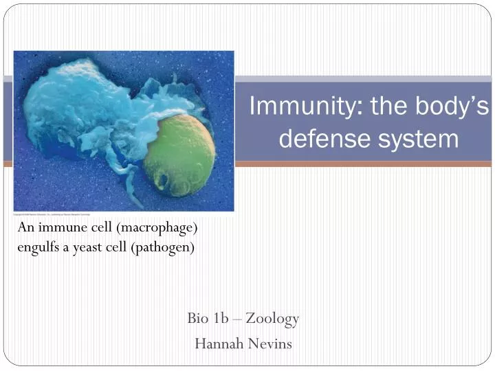
Immunity: the body’s defense system. Bio 1b – Zoology Hannah Nevins. An immune cell (macrophage) engulfs a yeast cell (pathogen). The immune system recognizes foreign bodies and responds with the production of immune cells and proteins

E N D
Immunity: the body’s defense system Bio 1b – Zoology Hannah Nevins An immune cell (macrophage) engulfs a yeast cell (pathogen)
The immune system recognizes foreign bodies and responds with the production of immune cells and proteins Two strategies have evolved: the innate and the acquired immune systems Invaders : pathogens
Innate Immunity of Invertebrates • The digestive system is protected by low pH and an enzyme that digests microbial cell walls called lysosome • Hemocytes circulate within hemolymph and carry out phagocytosis, the ingestion and digestion of foreign substances including bacteria • In insects, an exoskeleton made of chitin forms the first barrier to pathogens
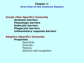
Innate Immunity of Vertebrates • Innate defenses include: • barrier defenses, phagocytosis, antimicrobial peptides • Additional defenses are unique to vertebrates: the inflammatory response and natural killer cells • The immune system of mammals is the best understood of the vertebrates
Human Lymphatic system Interstitial fluid Adenoid Tonsil Blood capillary Lymph nodes Spleen Lymphatic vessel Tissue cells Peyer’s patches (small intestine) Appendix Lymphatic vessels Lymph node Masses of defensive cells Fig. 43-7
Barrier Defenses • Mucustraps and allows for the removal of microbes • Many body fluids including saliva, mucus, and tears are hostile to microbes • The low pH of skin and the digestive system prevents growth of microbes • Barrier defenses include the skin and mucous membranes of the respiratory, urinary, and reproductive tracts
Cellular Innate Defenses • White blood cells (leukocytes) engulf pathogens in the body • Groups of pathogens are recognized by Toll-like receptors (TLR)
Phagocytosis:(=eating, =cells)engulfing pathogens Microbes PHAGOCYTIC CELL Vacuole Lysosome containing enzymes Exocytosis – cellular debris is released Fig. 43-3
Phagocytosis • A white blood cell engulfs a microbe, then fuses with a lysosome to destroy the microbe • There are different types of phagocytic cells: • Neutrophils engulf and destroy microbes • Macrophages are part of the lymphatic system and are found throughout the body • Eosinophils discharge destructive enzymes • Dendritic cells stimulate development of acquired immunity
How your skin keeps out pathogens • Ruptured mast cells (in tissue) release histamines chemical signal to other phagocytic cells • Capillaries dilate, • increase blood flow • increase phagocytic cells • The clotting process also starts • Platelets • Clotting factors signal • Fibrin produced Pathogen Splinter Chemical signals Macrophage Mast cell Capillary Red blood cells Phagocytic cell Fig. 43-8-1
Fig. 43-8-2 Pathogen Splinter Chemical signals Macrophage Fluid Mast cell Capillary Red blood cells Phagocytic cell
Fig. 43-8-3 Pathogen Splinter Chemical signals Macrophage Fluid Mast cell Capillary Phagocytosis Red blood cells Phagocytic cell • More phagocytic cells are released • Pathogenic bacteria are engulfed and destroyed • Pus, a fluid rich in white blood cells, dead microbes, and cell debris, accumulates at the site of inflammation
Lymphocyte maturation Thymus • White blood cells called lymphocytes recognize and respond to antigens, foreign molecules • Lymphocytes that mature in the thymus above the heart are called T cells,and those that mature in bone marrow are called B cells Lymph nodes Spleen Lymphatic vessels Fig. 43-7
Acquired Immunity results from B- and T-cells • T-cells • Thymus • Combats viruses (intracellular pathogens) • B-cells • Bone marrow & spleen • Combats bacteria (extracellular pathogens)
Pathogens have antigens, B-cells have antibodies • Antigens: • Each pathogen type has unique surface molecules • Antibody binding: • Causes antibodies to be secreted from B-cell • Antibodies: • Surface proteins of B-cell • Match antigens
Both B- and T-cells have Antigen binding sites Antigen- binding site Antigen- binding site Antigen- binding site Disulfide bridge V V V V Variable regions V V C C Constant regions C C C C Light chain Transmembrane region Plasma membrane chain chain Heavy chains Disulfide bridge B cell Cytoplasm of B cell Cytoplasm of T cell T cell (a) B cell receptor (b) T cell receptor Fig. 43-9
Antigen- binding sites Epitopes (antigenic determinants) Antigen-binding sites Antigen Antibody A Antibody C V V V V C C C C Antibody B Fig. 43-10
Lymphocyte Development • The acquired immune system has three important properties: • Receptor diversity • A lack of reactivity against host cells • Immunological memory
A Pathogen is tagged for Attack; a B-cell is “selected for cloning • Antibodies cause: • Neutralization • Agglutination • Precipitation • rupture • Selection causes rapid clonal replication Selection Replication
Fig. 43-14 Antigen molecules B cells that differ in antigen specificity Antigen receptor Antibody molecules Clone of memory cells Clone of plasma cells
The B-cells form Two cell Types: • Memory Cells • Long-lived • Await future encounters with specific antigen • Plasma Cells • Secrete many antibodies to mark and block more bacteria Selection Replication
Secondary Immune Response Primary immune response Secondary immune response • Get a disease, you get natural immunization • e.g. chicken pox • Immunization: injecting chemical or heat inactivated antigens • a.k.a vaccination 104 103 Antibody concentration Antibodies to A Antibodies to B 102 101 100 0 7 14 21 28 35 42 49 56 Exposure to antigen A Exposure to antigens A and B Time (days)
Fig. 43-15 Primary immune response to antigen A produces antibodies to A. Secondary immune response to antigen A produces antibodies to A; primary immune response to antigen B produces antibodies to B. 104 103 Antibody concentration (arbitrary units) Antibodies to A Antibodies to B 102 101 100 0 7 14 21 28 35 42 49 56 Exposure to antigen A Exposure to antigens A and B Time (days)
Pathogens can evolve to avoid detection • Some pathogens change surface proteins • Memory cells can not recognize • Pathogens have shorter generation time relative to host, :. they can evolve faster • What does this mean for the efficacy of any given human-made antibiotic? • Some pathogens like AIDS hide inside your body’s cells • Intracellular invaders are dealt with by T-cells
Like B-cells, T-cells have… Microbe Antigen- presenting cell Infected cell Antigen associates with MHC molecule 1 • Diverse antigen receptors • Two types: Cytotoxic T-cell, Helper T-cell Antigen fragment Antigen fragment 1 1 Class I MHC molecule Class II MHC molecule 2 2 T cell receptor T cell receptor 2 T cell recognizes combination (a) Cytotoxic T cell (b) Helper T cell Fig. 43-12
Cytotoxic T-cells Cytotoxic T cell Once bound to CD8 receptor, T-cell becomes an “active killer” Perforin Granzymes CD8 TCR Class I MHC molecule Target cell Peptide antigen Fig. 43-18-1
Cytotoxic T-cells Cytotoxic T cell Perforin Granzymes CD8 TCR Class I MHC molecule Pore Target cell Peptide antigen Perforins – create pores in surface of target cell Granzymes – enter cell initiate apoptosis (cell death) Fig. 43-18-2
Cytotoxic T-cells Released cytotoxic T cell Cytotoxic T cell Perforin Granzymes CD8 TCR Dying target cell Class I MHC molecule Pore Target cell Peptide antigen Perforins – create pores in surface of target cell Granzymes – initiate apoptosis (cell death) Fig. 43-18-3
Fig. 43-17 Antigen- presenting cell Peptide antigen Bacterium Class II MHC molecule CD4 TCR (T cell receptor) Helper T cell + Cytokines Humoral immunity (secretion of antibodies by plasma cells) + Cell-mediated immunity (attack on infected cells) + + B cell Cytotoxic T cell
Cytotoxic T-cells attack diseased of cancerous cells labeled with MHCs • Normal cells make MHC (Major Histocompatibility Complex) molecules • Abnormal cells –like those with viruses – make MHCs which bind to viral proteins • Those antigens are presented on the surface of the infected cell • Then detected by cytotoxic T-cells … and the infected cell is destroyed
Major Histocompatibility Complex Genes have ~100 Alternative Alleles • Each MHC type presents a different type of antigen for T-cells to recognize as alien • Gene polymorphism increases chances of matching antigens • Thus increased MHC diversity = increased disease resistance • One study looked at male selection using old t-shirts and MHC analysis: females favor males with MHCs which differ from their own --- why is this adaptive?